Latest Science Note
Dojindo Science Note [July 5, 2023]
|
Scientists have unveiled that in comparison to young mice, one-third of old microglia show Lipofustin-related autofluorescence (AF), characterized by profound changes in lipid and iron content, phagocytic activity, and oxidative stress. Pharmacological removal of microglia in older mice successfully eliminated AF-microglia, following the repopulation of new functional microglia leads to an improvement in age-related neurological impairments and reduces neurodegeneration after traumatic brain injury. Learn more about how the authors phenotyped AF-microglia using Lipi-Blue* for Lipid droplet labeling, and FerroOrange* for iron labeling. (Please refer to Fig. 1E, 5F, 9E for FerrOrange, Fig. 5D, 7E, for Lipi-Blue) |
|||
|
Brain injury accelerates the onset of a reversible age-related microglial phenotype associated with inflammatory neurodegeneration Click here for the original article: Rodney M Ritzel, et. al., Sci Adv (2023) Point of Interest |
|||
| Related Techniques | |||
| Lipid droplets detection | Lipi-Blue / Green / Red / Deep Red | ||
| Intracellular ferrous ion (Fe2+) detection | FerroOrange | ||
| Mitochondria ferrous ion (Fe2+) detection | Mito-FerroGreen | ||
| Lysosomal function assay | Lysosomal Acidic pH Detection Kit-Green/Deep Red NEW | ||
| Lysosomal Acidic pH Detection Kit-Green/Red | |||
| Cellular senescence detection (Live cell imaging or FCM) | Cellular Senescence Detection Kit | ||
| Cellular senescence detection (Plate reader) | Cellular Senescence Plate Assay Kit | ||
| Mitochondrial superoxide detection | MitoBright ROS Deep Red - Mitochondrial Superoxide Detection | ||
| Total ROS detection | Highly sensitive DCFH-DA or Photo-oxidation Resistant DCFH-DA | ||
| Related Applications | |||
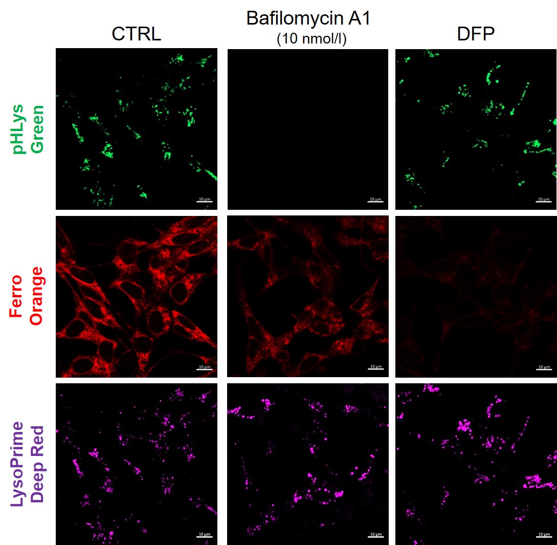
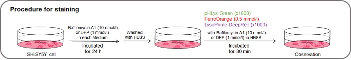
Measurement of intracellular iron changes and lysosomal pH changes
|
|||