CFSE
Fluorescent Dye for Cytoplasmic Staining
-
Product codeC309 CFSE
-
CAS No.150347-59-4
-
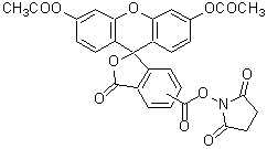
Chemical name5- or 6-(N-Succinimidyloxycarbonyl)fluorescein 3',6'-diacetate
-
MWC29H19NO11=557.46
| Unit size | Price | Item Code |
|---|---|---|
| 10 mg | Find your distributors | C309-08 |
Description
CFSE is cell-membrane permeable and readily accumulates inside viable cells where it covalently attaches to intracellular proteins (Fig. 1). Hydrolyzed CFSE emits fluorescence and covalently attached fluorescein molecules do not leak from cells. CFSE-labeled cells can be monitored over several weeks in vivo. Therefore, CFSE is utilized for detection of viable cell as well as for the long-term observation of cell activities by fluorescent microscopy. The excitation and emission wavelengths of CFSE-labeled cells are 500 nm and 520 nm, respectively. CFSE-stained cells are shown in Fig. 2.
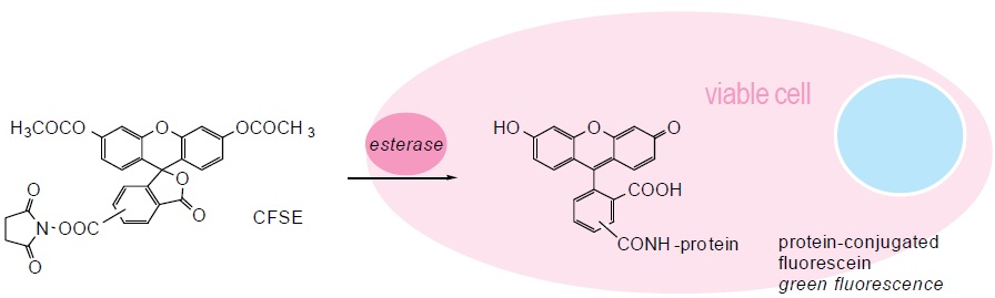
Fig. 1 Cell staining mechanism
Technical info
1. Prepare 1 mM CFSE solution with DMSO. Dilute it to prepare 10-50 μM CFSE solution with PBS or an appropriate buffer.
2. Add CFSE solution with 1/10 of the volume of cell culture medium to the cell culture.
3. Incubate the cell at 37ºC for 15 to 30 min.
4. Wash cells twice with PBS or an appropriate buffer.
5. Observe the cells under a fluorescence microscope with 490 nm excitation and 530 nm emission filters.
References
1) M. Bronner-Fraser, "Alterations in Neural Crest Migration by a Monoclonal Antibody That Affects Cell Adhesion", J. Cell Biol., 1985, 101, 610.
2) A. Nose and M. Takeichi, "A Novel Cadherin Cell Adhesion Molecule: Its Expression Patterns Associated With Implantation and Organogenesis of Mouse Embryos", J. Cell Biol., 1986, 103, 2649.
3) S. A. Weston and C. R. Parish, "New Fluorescent Dyes for Lymphocyte Migration Studies Analysis by Flow Cytometry and Fluorescent Microscopy", J. Immunol. Methods, 1990, 133, 87.
4) C. K. Raymond, P. J. O'hara, G. Eichinger, J. H. Rothman and T. H. Stevens, "Molecular Analysis of the Yeast VPS3 Gene and the Role of Its Product in Vacuolar Protein Sorting and Vacuolar Segregation during the Cell Cycle", J. Cell Biol., 1990, 111, 877.
5) G. Radcliff, R. Waite, J. Lefevre, M. D. Poulik and D. M. Callewaert, "Quantification of Effector/Target Conjugation Involving Natural Killer(NK) or Lymphokine Activated Killer(LAK) Cells by Two-color Flow Cytometry", J. Immunol. Methods, 1991, 139, 281.
6) S. A. Weston and C. R. Parish, "Calcein: a Novel Marker for Lymphocytes Which Enter Lymph Nodes", Cytometry, 1992, 13, 739.
7) L. S. D. Clerck, C. H. Bridts, A. M. Mertens, M. M. Moens and W. J. Stevens, "Use of Fluorescent Dyes in the Determination of Adherence of Human Leucocytes to Endothelial Cells and the Effects of Fluorochromes on Cellular Function", J. Immunol. Methods, 1994, 172, 115.
Handling and storage condition
| Appearance: | White to slightly yellow powder |
|---|---|
| Purity (HPLC): | ≧ 95.0 % |
| Solubility in Dimethyl sulfoxide: | To pass test |
| -20°C |
Related products
The following related products are also used in research related to this product.