Systemic Signals in Aging and Rejuvenation [May 14, 2025]

Previous Science Note

Cellular senescence spreads through tissues via secreted factors, contributing to tissue dysfunction, chronic inflammation, and impaired regeneration. This Science Note introduces recent insights into the in vivo spread of senescence and highlights emerging evidence that rejuvenating factors can also spread, suggesting new avenues for modulating aging and restoring tissue function.

Mitochondrial DNA released by senescent tumor cells enhances PMN-MDSC-driven immunosuppression through the cGAS-STING pathway (Immunity, 2025)
Summary: Senescent cells, including those undergoing therapy-induced senescence, actively shed mitochondrial DNA (mtDNA) into the tumour microenvironment via extracellular vesicles. This mtDNA is taken up by polymorphonuclear myeloid-derived suppressor cells, enhancing their immunosuppressive activity through cGAS-STING-NF-κB signalling and promoting tumour progression.

Highlighted technique: To assess mtDNA transfer from tumour senescent cells, extracellular vesicles (EVs) were isolated and confirmed to be mtDNA enriched. Their uptake by BM-MDSCs led to intracellular mtDNA accumulation, whereas EV-suppressed senescent cells failed to induce mtDNA accumulation during co-culture with BM-MDSCs.

 Related technique  Cellular Senescence Detection, Exosome Labeling Dye

Hepatocellular senescence induces multi-organ senescence and dysfunction via TGFβ (Nature Cell Biology, 2024)
Summary: Hepatocyte senescence in acute severe liver disease induces senescence in extrahepatic organs, contributing to multi-organ failure. This systemic spread is mediated by the TGFβ pathway and its inhibition can prevent secondary organ dysfunction.

Highlighted technique: his study used a mouse model enabling liver-specific and time-controlled deletion of MDM2, a suppressor of p53. MDM2 removal led to p53 accumulation and p21 upregulation in liver cells, allowing investigation of how liver senescence affects other organs.

 Related technique  Cellular Senescence Detection

Small extracellular vesicles from young plasma reverse age-related functional declines by improving mitochondrial energy metabolism (Nature Aging, 2024)
Summary: Small extracellular vesicles (sEVs) from young mice reverse age-related changes in aged mice by improving molecular, cellular and physiological functions, including lifespan extension and reduced senescence. These effects are mediated by miRNA, which improves mitochondrial function and metabolic processes in aged tissues.

Highlighted technique: In this study, researchers collected sEVs from the blood plasma of young mice and injected them into older mice to observe the effects. They examined lifespan, cognitive function and mitochondrial activity, which is often impaired in ageing, to show the reversal of signs of ageing in the mice.

 Related technique  Cellular Senescence Detection, Mitochondrial membrane potential detection

Previous Science Note

Related Techniques (click to open/close)
Target Kit & Probes
Cellular senescence detection SPiDER-βGal for live-cell imaging or flow cytometry / microplate reader / tissue samples.
Blue cellular senescence detection dye for fixed cells,  SPiDER Blue
Exosome Monitoring ExoSparkler Exosome Membrane Labeling Kit-Green / Red / Deep Red
Exosome Isolation ExoIsolator Exosome Isolation Kit and Isolation Filter
Mitochondrial membrane potential detection JC-1 MitoMP Detection Kit, MT-1 MitoMP Detection Kit
Mitochondrial Staining MitoBright LT Green / Red / Deep Red
Oxygen Consumption Rate(OCR) Detection Extracellular OCR Plate Assay Kit
Apoptosis detection in multiple samples Annexin V Apoptosis Plate Assay Kit
Cell proliferation/ cytotoxicity assay Cell Counting Kit-8 and Cytotoxicity LDH Assay Kit-WST
 Application Note I  (click to open/close)
  > Senescent Cells Lose Mitochondrial Activity

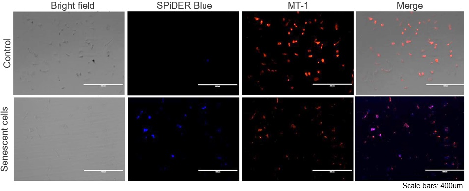
The senescent cell detection dye SPiDER Blue (SG07) and the mitochondrial membrane potential (MMP) dye MT-1 (MT13) were used to stain human microglial cells. Microscopy revealed that, compared to control cells, senescence-induced cells showed reduced MMP and increased SPiDER Blue fluorescence, reflecting elevated SA-β-Gal activity.

*This data was kindly provided by Dr. Supriya D. Mahajan, Department of Medicine, Jacobs School of Medicine & Biomedical Sciences.

1. Seed human microglia cells into a dish and incubate in an incubator set at 37 ℃ and equilibrated with 95% air and 5% CO2.
2. Dilute the MT-1 Dye (1:1000) in the cell culture medium.
3. Add MT-1 working solution to cells.
4. Incubate the cells for 30 minutes in an incubator set at 37 ℃ and equilibrated with 95% air and 5% CO2.
5. Discard the supernatant and wash the cells with HBSS twice.
6. Add 4% PFA/ PBS solution to the cells and incubate at room temperature for 30 minutes.
7. Discard the 4% PFA / PBS solution and wash the cells with PBS.
8. Add 15 µmol/l Spider Blue working solution and incubate at 37°C for 30 minutes.
9. Remove the working solution, and wash the cells with PBS.
10. Add Imaging Buffer solution and observe the cells under a fluorescence microscope.

 Application Note II (click to open/close)
  > Increased Senescence in Aged Adipose Tissue

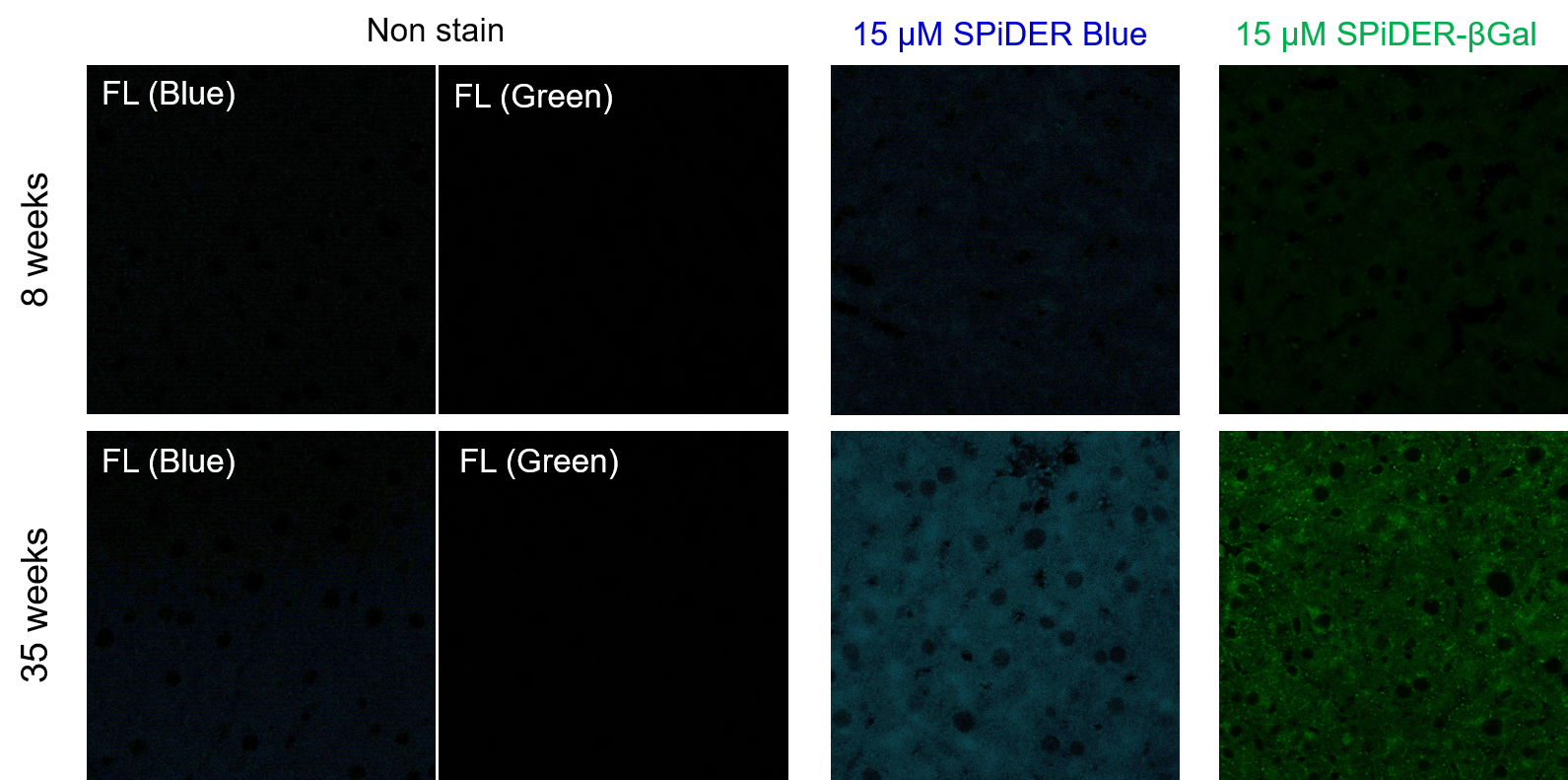
Frozen liver adipose tissue sections from 8-week-old and 35-week-old mice were stained with senescence detection probes SPiDER Blue (SG07) and SPiDER-βGal (SG02). Confocal microscopy revealed a marked increase in fluorescence intensity only in the 35-week-old samples, indicating an age-associated accumulation of senescent cells in older tissue.

1. 8-week-old and 35-week-old mouse liver adipose tissue (frozen sections) samples were prepared on glass slides. 
2. After washing once with PBS, 200 µl of 4% paraformaldehyde (PFA)/PBS solution was added and fixed at room temperature for 30 minutes. 
3. The supernatant was removed and washed once with PBS. 
4. Add 200 µl of 15 µM SPiDER Blue and 15 µM SPiDER-βGal prepared in Assay buffer and incubate at 37°C for 2 hours. 
5. The supernatant was removed and washed once with PBS. 
6. Add 1 drop of encapsulant (ProLong Glass Antifade Mountant, Thermo) and encapsulate with cover glass. 
7. Observed under a confocal laser microscope (40x magnification).

[Detection conditions]
SPiDER Blue: 405 nm (Ex), 400–500 nm (Em), 2.0%, 700V
SPiDER-βGal: 488 nm (Ex), 500–600 nm (Em), 1.0%, 600V


Product Classification

Product Classification