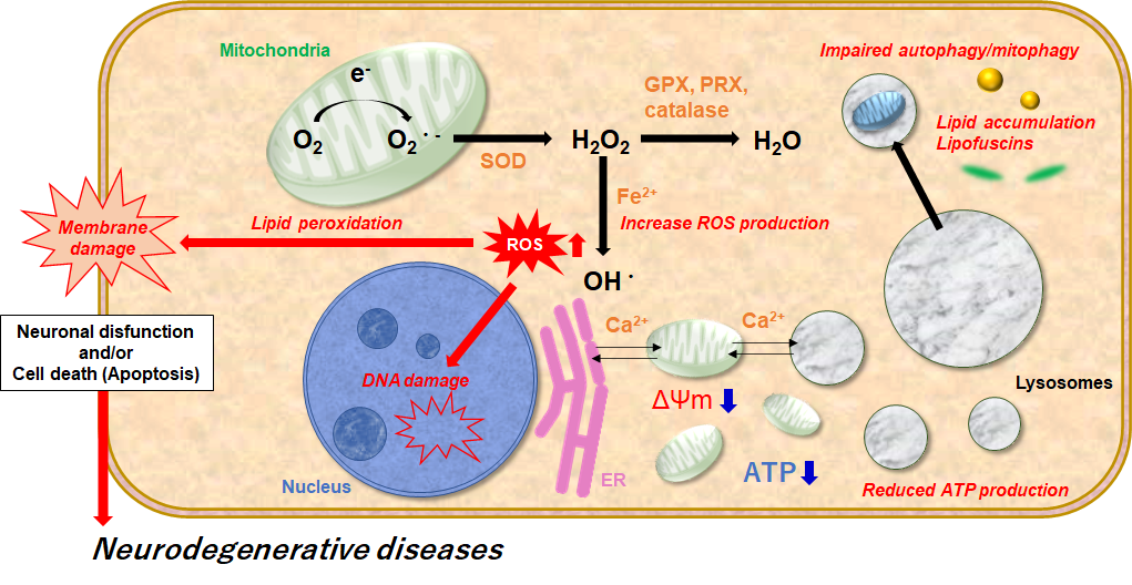
Mitochondria and lysosome dysfunction are particularly relevant in neurons, which strongly depend on oxidative phosphorylation for ATP production and on regulation of intracellular calcium levels for neuronal signaling. Mitochondrial dysfunction also has an impact on lysosomal function, suggesting a functional relationship between these two organelles. Lysosomal dysfunction is associated with impaired autophagy, dysregulation of mitochondrial quality control pathways, increased ROS production, and the accumulation of damaged mitochondria. 1), 2)
In this short review, we show how Dojindo’s products can be used to assess mitochondria and lysosome function for neurodegenerative disease research.
References
1. Nicoletta, P. et al., “Mitochondrial Dysfunction and Neurodegeneration in Lysosomal Storage Disorders”, Trends Mol Med., 2017, 23(2), 116-134.
2. Sana, O. et al., “Use of Hydrogen Peroxide and Peroxyl Radicals to Induce Oxidative Stress in Neuronal Cells”, Reviews in Agricultural Science, 2015, 3, 40-45.
Multiple Staining with Other Mitochondrial Markers
Decreased mitochondrial activity and mitochondrial dysfunction correlate with neurodegenerative diseases such as Alzheimer’s and Parkinson’s disease. More specifically, mitochondrial mass (MM), mitochondrial membrane potential (MMP), and mitochondrial ROS (mtROS) are all important and promising targets for mitochondria-related diseases. These mitochondrial attributes are dynamic and therefore require simultaneous analysis via multiple staining in a single sample.
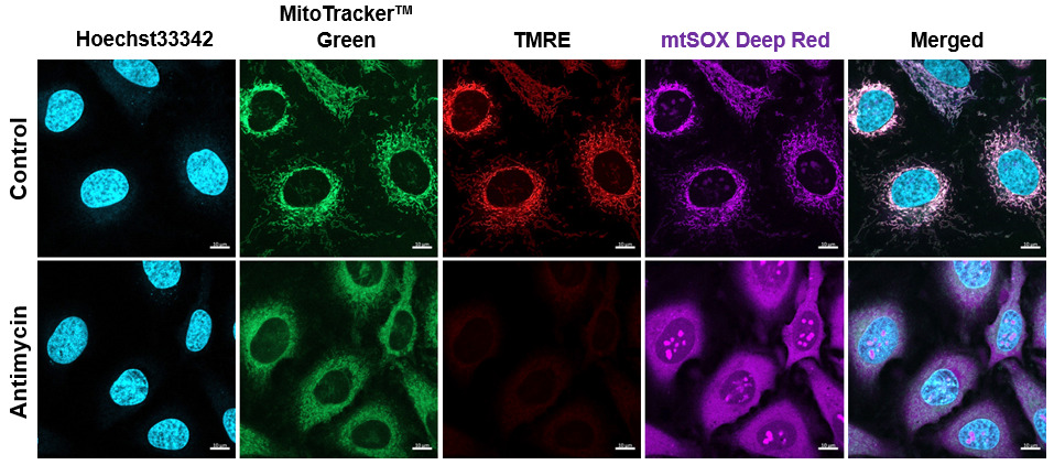
Blue : Nuclear stain: Hoechst33342 (Ex: 405 nm, Em: 450–495 nm)
Green : Mitochondrial mass stain: MitoTracker™ Green FM (Ex: 488 nm, Em: 500–550 nm)
Red : Mitochondrial membrane potential stain: TMRE (Ex: 561 nm, Em: 560–620 nm)
Purple : Mitochondrial superoxide stain: mtSOX Deep Red (Ex: 633 nm, Em 640–700 nm)
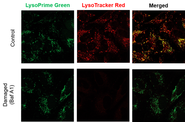
Observe Changes in Lysosomal Count and Function
Recent findings reveal that lysosomal dysfunction is related to some neurodegenerative disorders. Consequently, investigation of lysosomal function is attracting considerable interest in the scientific community. Existing lysosome probes are highly reliant on lysosomal pH; their emission intensity is unstable and varies significantly due to differences in acidity among lysosomes within a cell. This means it is difficult to understand if fluorescence changes are due to changes in lysosome count or function (pH).
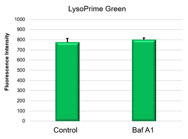
Dojindo’s LysoPrime Green, however, is not affected by variations in lysosome acidity that can occur after staining. Logically, lysosome count and function can therefore be evaluated by co-staining with LysoPrime Green and LysoTracker Red.
Detection Condition
Control : Normal condition, Bafilomycin A1: Inhibition of lysosomal acidification
LysoPrime Green filter sets : 488 nm (Ex), 500 – 570 nm (Em)
LysoTracker Red filter sets : 548 nm (Ex), 550 – 650 nm (Em)
Crosstalk between mitochondria and lysosomes plays an important role in neurodegenerative disease.
In this experiment, we simultaneously stained mitochondrial mass (MitoTracker™ Green), mitochondrial ROS (mtSOX Deep Red), mitochondrial membrane potential (TMRE), lysosomal content (LysoPrime Green), and lysosomal pH (LysoTracker™ Red) in neuron cells (SHSY-5Y). We then observed changes in lysosomes and mitochondria when treated with CCCP, Antimycin, and Oligomycin A, which are mitochondrial ROS inducers.
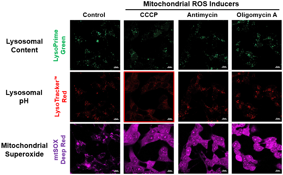
It is indicated that CCCP treatment not only changes mitochondrial ROS but also changes Lysosomal pH.
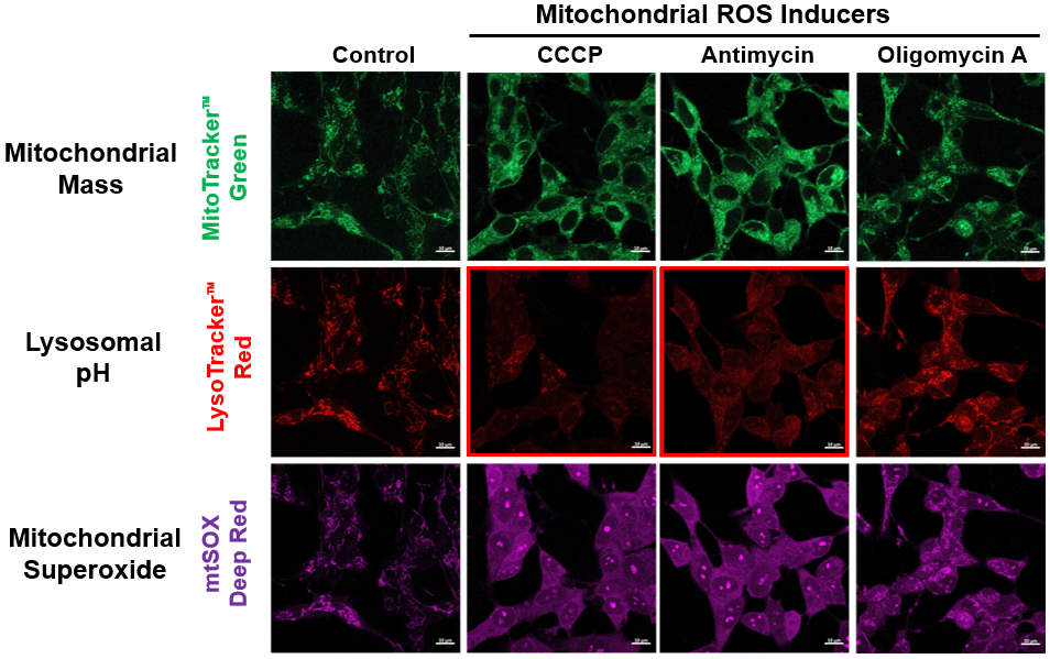
It is indicated that CCCP or Antimycin treatment changes mitochondrial membrane potential.
Related Products