Molecular Control of Mitochondrial Ca2+ Efflux in Neurons [Mar. 18, 2026] 

Mitochondrial Ca2+ control in neurons links activity to metabolism and is important for neuronal function and memory. Defining the proteins that regulate mitochondrial Ca2+ flux is therefore important for understanding cognitive decline and neurodegeneration. Recent studies showed that loss of the mitochondrial H+/Ca2+ exchanger Letm1 reduces Ca2+ efflux, thereby sustaining mitochondrial Ca2+ signals, extending metabolic activation, and enhancing long term memory in flies and mice. Another study showed that TMEM65 is necessary and sufficient for mitochondrial Na+/Ca2+ exchange, thereby identifying the molecular basis of mito-NCX, a major neuronal mitochondrial Ca2+ efflux pathway. Together, these findings clarify major regulators of neuronal mitochondrial Ca2+ efflux relevant to dementia related biology.

Mitochondrial Ca2+ efflux controls neuronal metabolism and long-term memory across species (Nature Metabolism, 2026)
Summary: Neuronal mitochondrial Ca2+ normally acts as a transient metabolic signal during activity, but whether its clearance influences long-term memory remains unclear. This study shows that Letm1, a mitochondrial H+/Ca2+ exchanger involved in Ca2+ efflux, prolongs metabolic activation when depleted, promotes long-term memory formation in flies and mice, and suggests relevance to memory decline in neurodegenerative disorders.

Highlighted technique: Pyruvate enters mitochondrial metabolism through the TCA cycle, so the authors measured pyruvate flux to test whether Letm1 depletion truly increases mitochondrial metabolism, not just ATP levels. They expressed Pyronic, a FRET-based pyruvate sensor, in rat hippocampal neurons, blocked mitochondrial metabolism with sodium azide, and used the rate of cytosolic pyruvate buildup as a readout of pyruvate flow into mitochondria.

TMEM65 functions as the mitochondrial Na+/Ca2+ exchanger (Nature Cell Biology, 2025)
Summary: Mitochondrial Na+/Ca2+ exchange (mito-NCX) is the main pathway for clearing mitochondrial Ca2+ in neurons, but its molecular identity has remained unresolved despite NCLX being widely viewed as the leading candidate. This study shows that TMEM65 is necessary and sufficient for mito-NCX, thereby strongly identifying TMEM65 as the exchanger itself and redefining the molecular basis of mitochondrial Ca2+ efflux relevant to neurodegeneration and dementia.

Highlighted technique: TThe authors evaluated mitochondrial NCX (mito-NCX) activity by treating HEK293 cells with altered TMEM65 expression with digitonin, which permeabilized the plasma membrane while preserving mitochondria, and then monitoring Na+-induced Ca2+ release into the external medium using Calcium Green-5N. They also isolated mitochondria from the cells and measured total mitochondrial Ca2+ by extracting Ca2+ with BAPTA under denaturing conditions in the presence of SDS.

Mitochondrial and Metabolic Activity Indicators (click to open/close)
Target Kit & Probes
Mitochondrial and Metabolic Activity Indicators Calcium Kit II - Fluo 4 and Calcium Kit II - Fura 2
Glycolysis/Oxidative phosphorylation Assay Glycolysis/OXPHOS Assay Kit
Oxygen consumption rate assay Extracellular OCR Plate Assay Kit
Intracellular ATP Assay ATP Assay Kit-Luminescence
Intact Mitochondria Fractionation IntactMito Fractionation Kit for Tissue
MitoComplex-I Activity Assay MitoComplex-I Activity Assay Kit
Mitochondrial Staining MitoBright LT Green / Red / Deep Red
Mitochondrial membrane potential detection JC-1 MitoMP Detection Kit, MT-1 MitoMP Detection Kit
Mitochondrial superoxide detection MitoBright ROS Deep Red - Mitochondrial Superoxide Detection
Application Note I (click to open/close)
  > Inhibition of Mitochondrial Electron Transport Chain


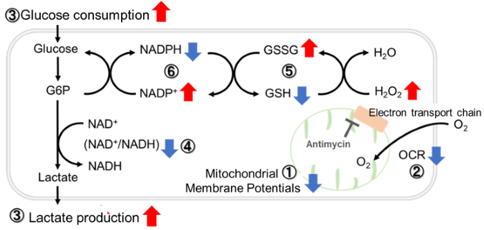
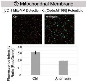
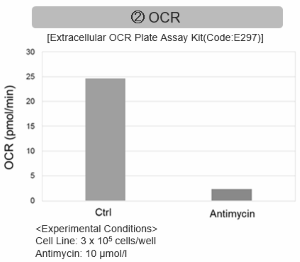
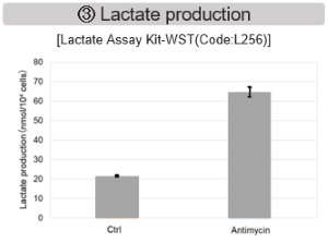
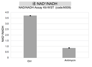
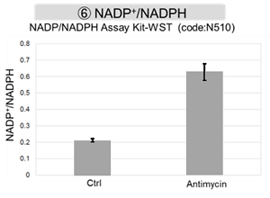
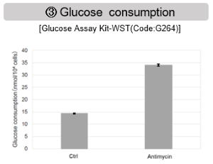
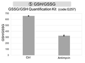
Antimycin stimulation of Jurkat cells was used to evaluate the changes in cellular state upon inhibition of the mitochondrial electron transport chain using a variety of indicators.

The results showed that inhibition of the electron transport chain resulted in (1) a decrease in mitochondrial membrane potential and (2) a decrease in OCR. In addition, (3) the NAD+/NADH ratio of the entire glycolytic pathway decreased due to increased metabolism of pyruvate to lactate to maintain the glycolytic pathway, (4) GSH depletion due to increased reactive oxygen species (ROS), and (6) increase in the NADP+/NADPH ratio due to decreased NADH required for glutathione biosynthesis were observed.


 Application Note II  (click to open/close)
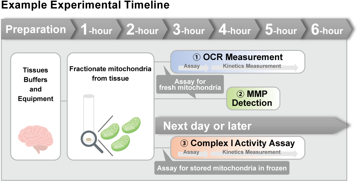
  > Activity Evaluation of Mitochondria Fractionated from Mouse Brain


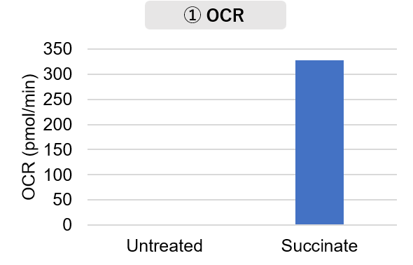
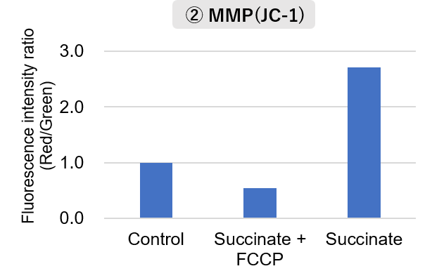
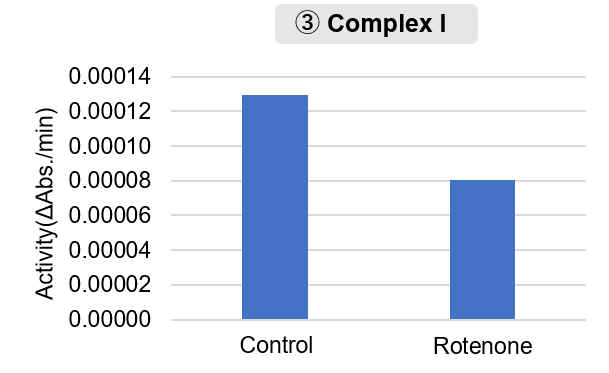
Mitochondria were isolated from mouse brain tissue, and oxygen consumption rate (OCR), mitochondrial membrane potential (MMP), and Complex I activity were measured.

The results showed that the addition of succinate, a substrate that activates Complex II of the electron transport chain, increased both OCR and MMP. In contrast, FCCP treatment reduced MMP, indicating that intact mitochondria were successfully fractionated.
Furthermore, in the Complex I activity assay, a decrease in activity was observed following treatment with rotenone, a Complex I inhibitor.

<Product used>
    Mitochondrial Fractionation: IntactMito Fractionation Kit for Tissue (Code: MT17)
 OCR measurement: Extracellular OCR Plate Assay Kit (Code: E297)
 MMP detection: JC-1 MitoMP Detection Kit (Code: MT09)
 Complex I activity assay:  MitoComplex- I Activity Assay Kit (Code: MT18)

<Experimental Conditions>
OCR Measurement   
    Amount of mitochondria: 50 μg/well (as protein levels)
    Succinate: 10 mmol/l
MMP Detection  
    Amount of mitochondria: 50 μg/well (as protein levels)
    Succinate: 10 mmol/l,   FCCP: 4 μmol/l
Complex I Activity Assay  
    Amount of mitochondria: 20 μg/well (as protein levels)
       Rotenone: 10 μmol/l


Previous Science Note

Product Classification

Product Classification