Neuronal Lipids and Disease-Related Dysfunction [Feb. 3, 2026] 

Neuronal lipids support mitochondrial energy production and membrane phospholipid composition, both important for neuronal function. Glucose has been positioned as the major energy source in neurons, yet recent work shows that triglycerides stored in presynaptic lipid droplets can serve as fuel under low glucose, and that inhibiting this utilization causes rapid hypothermia in mice. Because polyunsaturated fatty acids (PUFAs) can contribute to ferroptosis, PUFA lowering is sometimes discussed as protective, but an inherited ALS/FTD form shows reduced PUFA in neuronal membrane phospholipids, and boosting fatty acid desaturation to restore PUFA production improves neurodegeneration-related readouts. Together, these findings indicate that neuronal lipid utilization and membrane lipid composition can influence disease-relevant readouts.

Triglycerides are an important fuel reserve for synapse function in the brain (Nature Metabolism, 2025)
Summary: This study shows that neurons can use triglycerides stored in lipid droplets at presynaptic terminals as a readily mobilizable fuel to support mitochondrial energy production, especially during activity or when glucose is limited. Blocking triglyceride breakdown or mitochondrial fatty-acid entry leads to lipid droplet accumulation at synapses, failure of sustained synaptic vesicle recycling under glucose deprivation, and rapid hypothermia in mice, highlighting the importance of synaptic lipid-fuel flux for brain energy homeostasis.

Highlighted technique: To track the transfer of lipid droplet–associated fatty acids to mitochondria in cultured neurons, the authors performed a fluorescent fatty-acid tracer assay. Neurons were loaded with the fatty-acid analog BODIPY 558/568 C12 (Red-C12) under conditions that inhibit lipid droplet triglyceride breakdown, and mitochondria and lipid droplets were co-stained and imaged by confocal microscopy to confirm and quantify Red-C12 co-localization with mitochondria.

Neuronal polyunsaturated fatty acids are protective in ALS/FTD (Nature Neuroscience, 2025)
Summary: This study shows that, in a common inherited form of ALS/FTD, neuronal membrane phospholipids shift toward reduced polyunsaturated fatty acid (PUFA) content. Enhancing fatty-acid desaturation to restore PUFA production within neurons improved neurodegeneration-related readouts under experimental conditions, suggesting that reduced membrane polyunsaturation may contribute to neuronal vulnerability in some neurodegenerative settings.

Highlighted technique: To test whether increasing neuronal PUFA levels can rescue a disease-relevant cellular phenotype, the authors generated human spinal neurons from iPS cells derived from patients with a major inherited form of ALS/FTD and non-neurological controls, and boosted intracellular PUFA production by expressing fatty-acid desaturases. They then applied a defined glutamate excitotoxic stress and quantified phenotype changes by measuring cell death with a fluorescent dead-cell stain and cell survival using a standard viability assay.
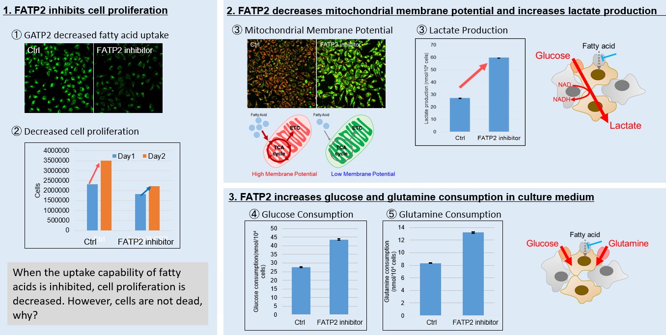
Previous Science Note

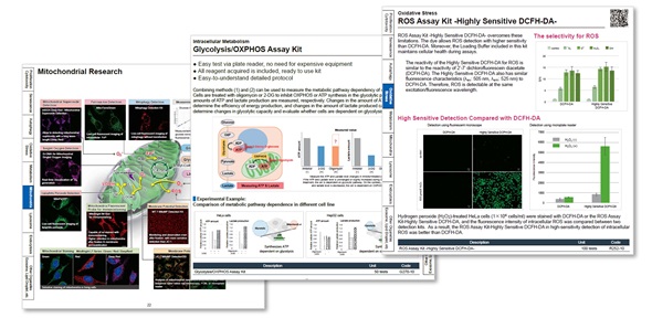
Metabolic Activity Assays (click to open/close)
Target Kit & Probes
Lipid Droplet Staining Lipi-Blue/ Green/ Red/ Deep Red
Fatty Acid Uptake Capacity Assay Fatty Acid Uptake Assay Kit
Mitochondrial Staining MitoBright LT Green / Red / Deep Red
Mitochondrial membrane potential detection JC-1 MitoMP Detection Kit, MT-1 MitoMP Detection Kit
Mitochondrial superoxide detection MitoBright ROS Deep Red - Mitochondrial Superoxide Detection
Intact Mitochondria Fractionation IntactMito Fractionation Kit for Tissue
MitoComplex-I Activity Assay MitoComplex-I Activity Assay Kit
Glycolysis/Oxidative phosphorylation Assay Glycolysis/OXPHOS Assay Kit
Oxygen consumption rate assay Extracellular OCR Plate Assay Kit
Application Note (click to open/close)
  > Fatty acid starvation induced by uptake inhibitor evoke reprogramming of cellular metabolism 


Mitochondrial fatty acid β-oxidation and oxidative phosphorylation (OXPHOS) are crucial biochemical processes that metabolize fats and sugars to produce ATP, the cell's primary energy source. In this section, we underscored the significance of fatty acid starvation and energy pathways, with an emphasis on the fatty acid uptake inhibitor, FATP2. Here are the key findings from our experiments conducted on HeLa cells:

Image 1: Inhibition of fatty acid uptake results in reduced cell proliferation, though it does not lead to cell death. This was determined through the use of a Cell Counting Kit-8 and Fatty Acid Uptake Kit.

Image 2: Fatty acid starvation shifts cellular metabolism from OXPHOS to glycolysis, as indicated by the Glycolysis/JC-1 MitoMP Assay Kit. 

Image 3: When fatty acid uptake is inhibited, a compensatory increase in glucose and glutamine uptake occurs to preserve cell viability, as observed using the Glucose Assay Kit and Glutamine Assay Kit.

Products in Use
for Fatty Acid Uptake Assay
  ① 
Fatty Acid Uptake Assay Kit

for Cell Proliferation/Cytotoxicity Assay
  ② 
Cell Counting Kit-8

for Glycolysis Assay and Mitochondrial Membrane Potential Detection
  ③
 Glycolysis/JC-1 MitoMP Assay Kit

for Glucose and Glutamine Consumption Assay
   Glucose Assay Kit

  ⑤ Glutamine Assay Kit

Previous Science Note  


Product Classification

Product Classification