Previous Science Note
|
Lysosomes and cell death pathways are fundamental to the regulation of cellular homeostasis, yet their dysregulation can contribute to pathological conditions. Recent studies highlight how impaired lysosomal function can trigger various forms of cell death. This Science Note introduces emerging insights into lysosomal dynamics and the mechanisms of cell death. |
||||||||||||||||||||||
|
Summary: The accumulation of lipid peroxidation within lysosomes increases lysosomal membrane permeability, leading to the release of iron into the cytosol and subsequently triggering ferroptosis. Increasing lysosomal membrane permeability may be a promising strategy to overcome ferroptosis resistance in certain cell types. Highlighted technique: In this study, lipid peroxide accumulation within lysosomes was visualised following induction of ferroptosis. In addition, increased lysosomal pH, cytosolic diffusion of internalised FITC-dextran and iron redistribution from lysosomes to the cytosol were observed, demonstrating that lysosomal membrane permeabilization contributes to ferroptosis. Related technique Accurate lysosomal detection / Fe2+ live-imaging (used in this article) / Lipid peroxide detection (used in this article) |
||||||||||||||||||||||
|
Summary: This study develops tNeurons, an aged human neuron model, and shows that lysosomal dysfunction in these cells drives amyloid β accumulation, inflammation and cell death, highlighting tNeurons as a powerful platform to study early pathogenic events and lysosomes as a therapeutic target in Alzheimer's disease. Highlighted technique: tNeurons are human neurons directly converted from aged dermal fibroblasts without transitioning through a pluripotent state, preserving age-associated dysfunctions such as lysosomal and mitochondrial impairments and disrupted proteostasis. They provide a valuable model for studying the progression of age-related diseases, including Alzheimer’s disease. Related technique Accurate lysosomal detection / Senescence assay / Apoptosis plate assay |
||||||||||||||||||||||
|
Summary: In Alzheimer's disease models, impaired lysosomal acidification causes Aβ and amyloid precursor protein-beta C-terminal fragment (APP-βCTF) accumulation in autolysosomes. This dysfunction leads to the formation of PANTHOS, a flower-like aggregation of autophagic vacuoles, along with lysosomal membrane permeabilization and cell death, promoting senile plaque development. Highlighted technique: In this study, neuronal autolysosomal acidification and dysfunction were assessed using a neuron-specific mRFP-eGFP-LC3 probe, where the pH-sensitive eGFP signal is quenched while the pH-stable mRFP signal persists in acidic compartments, enabling detection of acidification defects by multiplex confocal and correlative light-electron microscopy. Related technique Accurate lysosomal detection / Autophagic Flux Assay Kit |
||||||||||||||||||||||
All Related Techniques (click to open/close)
|
||||||||||||||||||||||
Application Note (click to open/close)
|
||||||||||||||||||||||
|
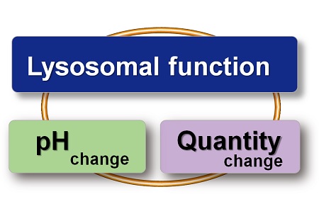
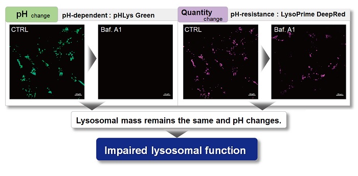
With existing reagents, it was difficult to determine whether lysosomal mass or their function (pH) fluctuated because the discussion was based on changes in the fluorescence brightness of a single dye. This kit contains pHLys Green, which is highly specific to lysosomes and shows pH-dependent changes in fluorescence, and pH-resistant LysoPrime Deep Red. Using these two dyes, lysosomal pH and volume of the same sample can be measured for a detailed analysis of lysosomal function.
|
|||
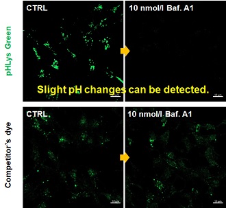
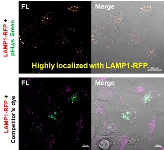
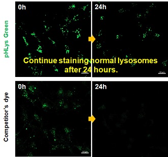
| Existing lysosomal pH detection reagents have issues with dye localization, pH sensitivity, and retention. pHLys Green is a dye that solves these issues. The improved dye retention and localization enable detection of normal lysosomes, and the improved pH sensitivity enables detection of slight pH changes. | |||
| 1. High sensitive pH detection Comparison of pH response of cells treated with low concentrations of lysosomal acidification inhibitor Bafilomycin A1 |
2. High specificity for lysosomes Comparison of specificity for lysosomes using lysosomal marker protein LAMP1-GFP expressing cells |
3. High retention in lysosomes Comparison of intracellular retention |
|
 |
 |
 |
|
|
Product in Use: Related Product: |
|||