
How Processing Fatty Acids Shapes Ferroptosis Sensitivity

Previous Science Note  

Fatty acids taken up by cells can be used for energy, stored as triglycerides in lipid droplets, or incorporated into membrane phospholipids. Because PUFA-containing phospholipids are readily oxidized, the metabolic destination of fatty acids can influence ferroptosis sensitivity.
The first paper showed that even when fatty acids caused similar levels of lipid droplet accumulation, their cis/trans double-bond geometry altered intracellular conversion and incorporation into membrane phospholipids, resulting in different ferroptosis responses. The second paper showed that human fibroblasts in an oncogene-induced senescence model increased triglyceride storage and PUFA-derived lipid mediator production, thereby reducing PUFA-containing membrane lipids and increasing ferroptosis resistance.
Together, these studies show that lipid droplet abundance alone does not indicate whether fatty acid accumulation protects cells from or sensitizes them to ferroptosis. Evaluating lipid droplets together with lipid peroxidation and cell death provides a clearer view of how intracellular fatty acid handling shapes ferroptosis sensitivity.

Ferroptosis Guide

Double-bond geometry determines fatty acid metabolic fate and ferroptosis sensitivity
(Harris et al., bioRxiv, 2026)

Summary
The researchers compared 62 structurally diverse fatty acids in two cancer cell lines. The trans PUFA linoelaidic acid increased ferroptosis sensitivity more strongly than cis linoleic acid, while the trans MUFA petroselaidic acid also increased sensitivity after enzymatic conversion into a PUFA. Linoelaidic acid increased oxidation-prone phospholipids containing two PUFA chains, whereas petroselaidic acid promoted oxidation-prone membrane lipids through a different metabolic route. Although all three fatty acids similarly increased lipid droplets and neutral lipids, blocking lipid droplet formation did not prevent sensitization. These findings show that, together with chain length and unsaturation, cis/trans double-bond geometry influences fatty acid metabolism and membrane incorporation, producing differences in ferroptosis sensitivity that are not explained by lipid droplet abundance.

Highlighted technique
To determine whether fatty acid structure, rather than lipid droplet abundance alone, influences ferroptosis sensitivity, the researchers used fluorescent probes to measure lipid droplets, lipid peroxidation, and cell death after ferroptosis induction. An antioxidant that suppresses ferroptosis confirmed the type of cell death, while blocking triglyceride synthesis tested whether lipid droplet formation accounted for differences in sensitivity.

Combining lipid droplet staining with measurements of lipid peroxidation, intracellular lipid radicals, and lipid peroxides can provide a multidimensional assessment of the relationship between lipid droplet accumulation and cellular lipid oxidation. 


Fatty acid channelling into triglycerides and oxylipins drives ferroptosis resistance during oncogenic BRAF-induced senescence
(Hess et al., Cell Death & Differentiation, 2026)

Summary
In this study, human fibroblasts induced into senescence accumulated lipid droplets and triglycerides, while PUFA-containing membrane phospholipids decreased. They also showed less lipid peroxidation and cell death following ferroptosis induction. Blocking triglyceride synthesis increased PUFA-containing membrane phospholipids and partially restored ferroptosis sensitivity. The senescent cells also showed increased production of PUFA-derived signaling lipids. Blocking both triglyceride synthesis and the production of these signaling lipids restored sensitivity to non-senescent levels. These results identify triglyceride synthesis and PUFA-derived signaling lipid production as two processes that reduce oxidation-prone membrane lipids and protect these senescent cells from ferroptosis.

Highlighted technique
To examine whether lipid storage contributes to ferroptosis resistance in senescent cells, the researchers confirmed senescence by measuring SA-β-gal activity and markers of cell-cycle arrest and SASP expression. Fluorescent probes were then used to evaluate lipid droplets, lipid peroxidation, and cell death. Blocking triglyceride synthesis reduced lipid droplet formation and increased lipid peroxidation and ferroptosis sensitivity.

Combining SA-β-gal detection with measurements of fatty acid uptake, lipid droplet accumulation, intracellular lipid radicals, lipid peroxides, and cell viability can provide a multidimensional view of how lipid storage and oxidative stress responses change during cellular senescence. 


Ferroptosis Related Indicators (click to open/close)
Target Kit & Probes
Lipid Droplet Staining Lipi-Blue/ Green/ Red/ Deep Red
Lipid Droplet Assay Blue/ Deep Red
Fatty Acid Uptake Capacity Assay Fatty Acid Uptake Assay Kit
Lipid Peroxidation Assay Lipid Peroxidation Probe -BDP 581/591 C11-
Lysosomal Lipid Radical detection
Lysosomal Lipid Radical Probe -Lyso-NBD-Pen-
Intracellular Lipid Radical detection Lipid Radical Probe -NBD-Pen-
Intracellular / mitochondrial lipid peroxidation detection Liperfluo, MitoPeDPP
Cell proliferation/ cytotoxicity assay Cell Counting Kit-8 and Cytotoxicity LDH Assay Kit-WST
Senescence-associated β-gal detection SPiDER-βGal for live-cell imaging or flow cytometry / microplate reader / tissue samples 
Blue cellular senescence detection dye for fixed cells,  SPiDER Blue
Intracellular / mitochondrial ferrous ion (Fe2+) detection FerroOrange, Mito-FerroGreen
Lysosomal ferrous ion (Fe2+) detection Lyso-FerroRed
Application Note  (click to open/close)
Experimental Example I: Hepatotoxicity test of drug-induced lipidosis using high-content imaging (click to open/close)

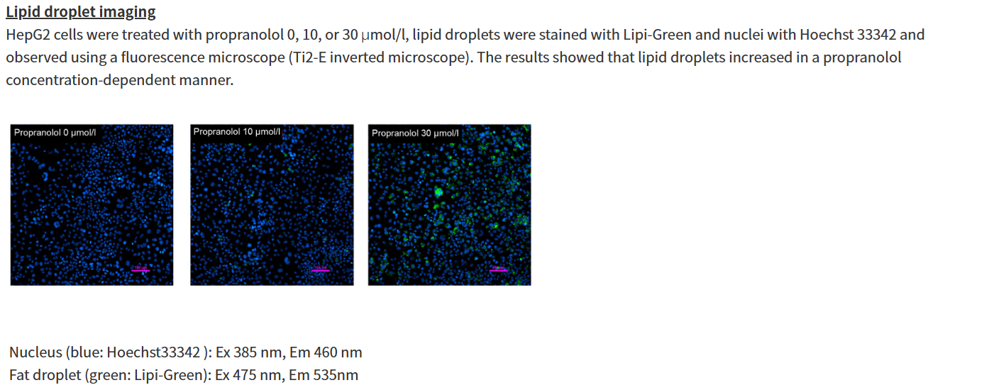
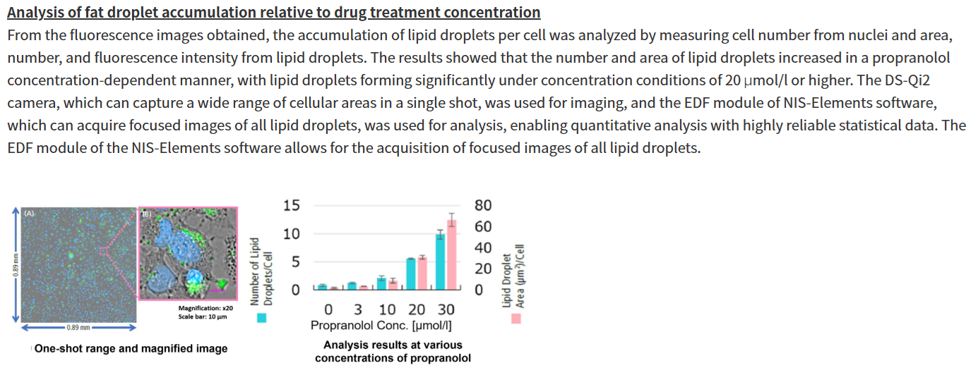
Propranolol (a sympathetic β-receptor blocker) was added to a human hepatocellular carcinoma cell line (HepG2 cells), and changes in lipid droplets were observed under a fluorescence microscope. The accumulation of lipid droplets was analyzed by measuring the number, area, and fluorescence intensity of lipid droplets from the acquired microscopic images.

High Content Analysis (HCA) microscope system
(Nikon Corporation https://www.microscope.healthcare.nikon.com/)

For details of staining and analysis methods, please refer to "APPLICATION NOTE: Hepatotoxicity test of drug-induced lipidosis using high-content imaging" by Nikon Corporation.


Experimental Example II: Changes in Each Indicator Following the Co-treatment of Ferroptosis Inducers and Lysosomal Inhibitors (click to open/close)

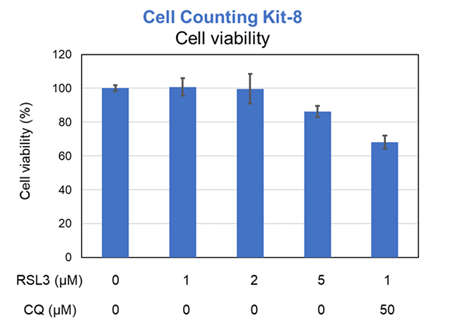
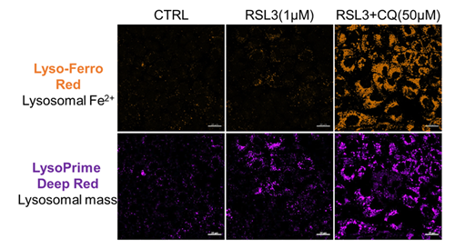
Previous studies have suggested that ferrotosis susceptibility varies among cancer cell lines. It has also been reported that increasing lysosomal stress in ferrotosis-resistant cancer cells can promote ferrotosis*. We treated A549 cells, which exhibit ferroptosis resistance, with the ferroptosis inducer RSL3 or RSL3 combined with the lysosomal inhibitor chloroquine (CQ) for 24 hours. Then, we analyzed changes in cell viability, lysosomal Fe²⁺, and lysosomal content. Treatment with RSL3 alone did not significantly alter cell viability or intracellular Fe²⁺ levels; however, some lysosomal aggregation (strong LysoPrime Deep Red signal) was observed. In contrast, cells treated with both RSL3 and CQ simultaneously exhibited increased intracellular Fe²⁺ levels, lysosomal enlargement, and decreased cell viability, which is consistent with previous reports. These results suggest that increased intracellular Fe²⁺ may promote ferroptosis.

* Saimoto. Y, et al., Nature Communications, 2025, 16, 3554.

[Products in use]
Cell viability: Cell Counting Kit-8 (Product code: CK04)
Lysosomal Fe2+: Lyso-FerroRed (Product code: L270)
Lysosomal mass: LysoPrime Deep Red - High Specificity and pH Resistance (Product code: L264)


Experimental Example III: Comparison of Lysosomal Lipid Radical Detection in Ferroptosis-Sensitive and -Resistant Cells (click to open/close)

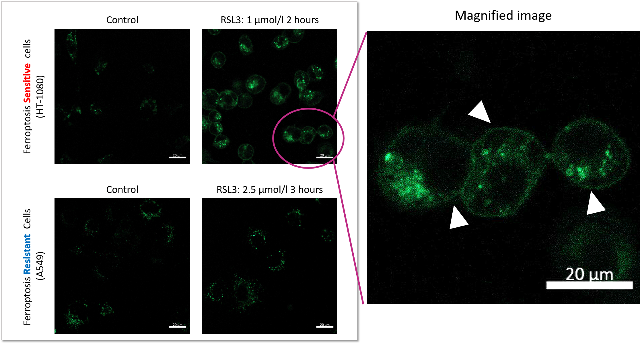
Changes in lysosomal lipid radicals were detected in HT-1080 cells, which are highly sensitive to ferroptosis, and A549 cells, which are resistant to ferroptosis, following treatment with RSL3, a ferroptosis inducer.

In A549 cells, a ferroptosis-resistant cell line, no significant difference in fluorescence intensity was observed compared with the control, even when the RSL3 treatment concentration and duration of RSL3 exposure were increased (concentration: up to 2.5 μmol/L; duration: up to 3 hours). In contrast, in HT-1080 cells, a ferroptosis-sensitive cell line, treatment with 1 μmol/L RSL3 for 2 hours increased fluorescence derived from lysosomal lipid radicals and caused changes in their localization.

These results demonstrate that this product can detect differences in lysosomal lipid radical generation and localization changes depending on the cell type.
Product Classification

Product Classification