



Science Note
|
Recent ferroptosis research increasingly focuses on where lipid radicals and lipid peroxidation occur inside cells. One study showed that lymph node metastatic melanoma weakens the GSH GPX4 axis while shifting toward FSP1 dependence, with FSP1 localizing around perinuclear lysosomes. Another study showed that lysosomal lipid peroxidation promotes membrane permeabilization, Fe²⁺ leakage, and broader lipid oxidation. Together, these findings suggest that lysosomal lipid oxidation may serve as an early readout for ferroptosis sensitivity and progression. |
 |
Summary:
Lymph node metastatic melanoma cells weaken the GSH GPX4 axis and become FSP1 dependent, with FSP1 enriched around perinuclear lysosomes, suggesting a link to lysosome proximal lipid oxidation control. FSP1 localized around perinuclear lysosomes, and FSP1 loss increased lipid oxidation under lysosome targeted ferroptosis stress. These findings suggest that lysosome proximal lipid oxidation control is important in lymph node metastatic melanoma.
Highlighted technique:
To assess how lymph node metastatic melanoma cells connect weakened GSH GPX4 defense with FSP1 localization, the authors measured GSH levels and lipid oxidation under ferroptosis stress, then examined FSP1 overlap with lysosome markers including LAMP1 by confocal imaging and lysosome enriched fractionation.
Lysosome-specific lipid radical detection by imaging supports analysis of localized lipid oxidation during ferroptosis. Combining this with pH-resistant and pH-dependent lysosome probes may enable evaluation of lysosomal changes associated with lipid oxidation and dysfunction.
Summary:
Ferroptosis is usually described as iron-dependent lipid peroxidation following GPX4 inhibition, but this study points to lysosomes as a likely early trigger site. In non-small cell lung cancer cells, lipid oxidation detected in lysosomes was linked to lysosomal membrane damage, iron leakage, broader lipid oxidation, and cell death. In low-susceptibility cells, this oxidative signal appeared without efficient membrane damage, suggesting that the transition from lysosomal oxidation to membrane permeabilization helps determine ferroptosis sensitivity.
Highlighted technique:
To assess whether lysosomal lipid oxidation progresses to membrane damage and iron release during ferroptosis, the authors measured lipid peroxidation, lysosomal pH changes, lysosomal ferrous iron, and cytosolic ferrous iron using fluorescence based imaging and flow cytometry. They also evaluated LDH release and cell viability to link these changes with ferroptosis.
Combining ferrous iron detection at both intracellular and lysosomal levels with LDH-based cytotoxicity measurement, enables evaluation of whether observed cell death is connected to ferroptosis and whether lysosomal iron dysregulation is the underlying cause.
).png)
).png)
Metabolic and Mitochondrial Activity Indicators (click to open/close)
| Target | Kit & Probes | ||||||
| Lysosomal ferrous ion (Fe2+) detection | Lyso-FerroRed | ||||||
| Intracellular / mitochondrial ferrous ion (Fe2+) detection | FerroOrange, Mito-FerroGreen | ||||||
| Intracellular / mitochondrial lipid peroxidation detection | Liperfluo, MitoPeDPPe | ||||||
| Lysosomal Lipid Radical detection |
Lysosomal Lipid Radical Probe -Lyso-NBD-Pen- | ||||||
| Intracellular Lipid Radical detection | Lipid Radical Probe -NBD-Pen- | ||||||
| Lipid Peroxidation Assay | Lipid Peroxidation Probe -BDP 581/591 C11- | ||||||
| Glutathione Quantification | GSSG/GSH Quantification Kit | ||||||
| Cell proliferation/ cytotoxicity assay | Cell Counting Kit-8 and Cytotoxicity LDH Assay Kit-WST | ||||||
| Lysosomal Function Analysis Kit | Lysosomal Acidic pH Detection Kit -Green/Red and Green/Deep Red | ||||||
| High Specific Lysosommal Detection | LysoPrime Green / Deep Red | ||||||
| Lysosomal Acidic pH Detection | pHLys Red | ||||||
Application Note I (click to open/close)
|
|||||||
|
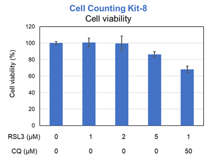
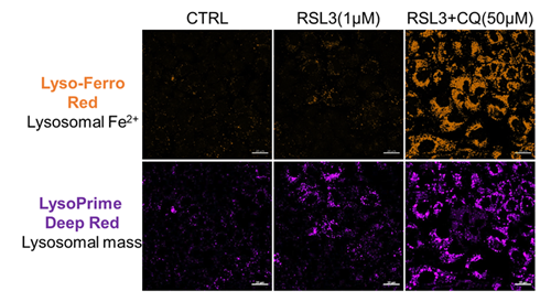
Previous studies have suggested that ferrotosis susceptibility varies among cancer cell lines. It has also been reported that increasing lysosomal stress in ferrotosis-resistant cancer cells can promote ferrotosis*. We treated A549 cells, which exhibit ferroptosis resistance, with the ferroptosis inducer RSL3 or RSL3 combined with the lysosomal inhibitor chloroquine (CQ) for 24 hours. Then, we analyzed changes in cell viability, lysosomal Fe²⁺, and lysosomal content. Treatment with RSL3 alone did not significantly alter cell viability or intracellular Fe²⁺ levels; however, some lysosomal aggregation (strong LysoPrime Deep Red signal) was observed. In contrast, cells treated with both RSL3 and CQ simultaneously exhibited increased intracellular Fe²⁺ levels, lysosomal enlargement, and decreased cell viability, which is consistent with previous reports. These results suggest that increased intracellular Fe²⁺ may promote ferroptosis. * Saimoto. Y, et al., Nature Communications, 2025, 16, 3554.
[Products in use] |
Application Note II (click to open/close)
> Comparison of Lysosomal Lipid Radical Detection in Ferroptosis-Sensitive and -Resistant Cells
|
Changes in lysosomal lipid radicals were detected in HT-1080 cells, which are highly sensitive to ferroptosis, and A549 cells, which are resistant to ferroptosis, following treatment with RSL3, a ferroptosis inducer. In A549 cells, a ferroptosis-resistant cell line, no significant difference in fluorescence intensity was observed compared with the control, even when the RSL3 treatment concentration and duration of RSL3 exposure were increased (concentration: up to 2.5 μmol/L; duration: up to 3 hours). In contrast, in HT-1080 cells, a ferroptosis-sensitive cell line, treatment with 1 μmol/L RSL3 for 2 hours increased fluorescence derived from lysosomal lipid radicals and caused changes in their localization. These results demonstrate that this product can detect differences in lysosomal lipid radical generation and localization changes depending on the cell type. 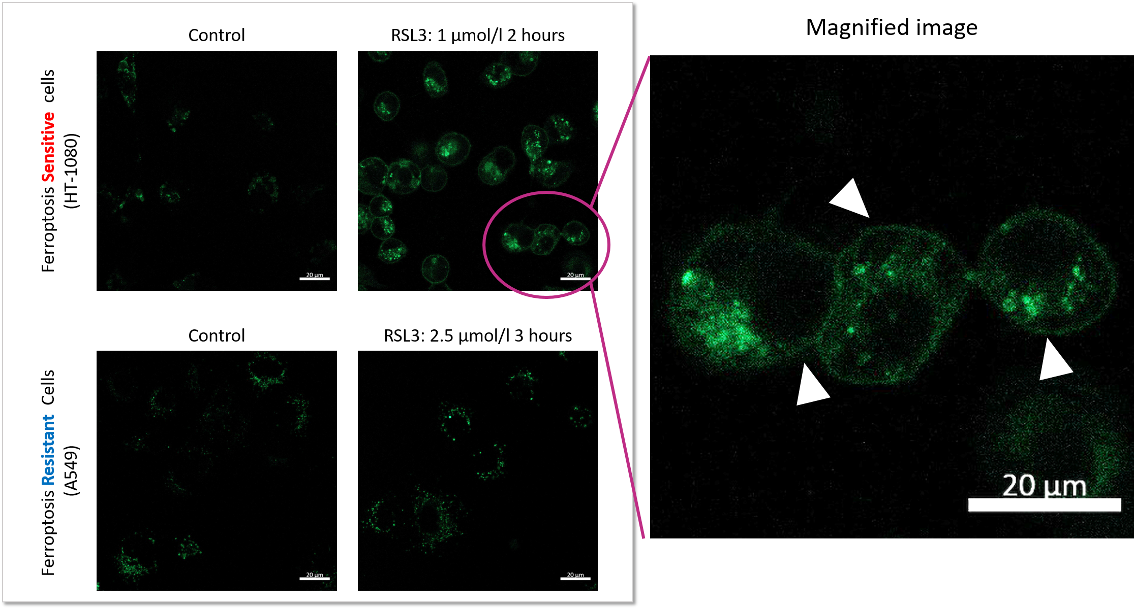 |
