Mitophagy Diversified: Discovery of a Microautophagy-like Mechanism [Jul. 15, 2025]


Autophagy is a cellular process in which intracellular components are degraded and recycled. It is broadly categorized into macroautophagy, which involves the formation of autophagosomes, and microautophagy, where lysosomes directly engulf target materials without autophagosome intermediates1. While mitophagy is typically mediated by macroautophagy, recent studies have uncovered a microautophagy-like mechanism by which lysosomes selectively remove damaged portions of mitochondria rather than the entire organelle2.

1. Autophagy genes in biology and disease (Nature Reviews Genetics, 2024)
Summary: This review provides an overview of the regulatory factors involved in autophagy, categorized from the perspectives of macroautophagy and microautophagy. It also summarizes mutations in autophagy-related genes associated with human diseases, particularly neurodegenerative disorders. These insights help clarify how autophagy dysfunction contributes to disease pathogenesis and whether a given disease may be linked to defects in autophagic pathways.

 Related techniques  Autophagic Flux Detection

2. Lysosomes drive the piecemeal removal of mitochondrial inner membrane (Nature, 2024)
Summary: Lysosomes maintain mitochondrial quality control not only by degrading the entire mitochondria but also by selectively removing damaged sections of the inner mitochondrial membrane (IMM). The damaged IMM protrudes through pores in the outer membrane formed by VDAC1, and nearby lysosomes engulf this IMM portion, leading to the formation of vesicles in the cytoplasm.

Highlighted technique: This study uses a combination of super-resolution microscopy, live-cell imaging, and specific fluorescent probes and proteins to visualize and track in real time the dynamic process of IMM protruding through the OMM and their subsequent uptake by lysosomes.

 Related techniques  Lysosomal Function Analysis, Mitophagy detection
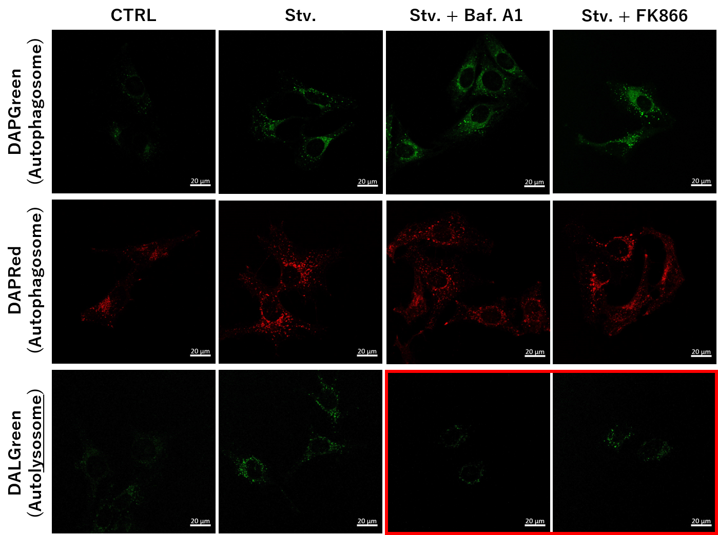
Previous Science Note

Related Techniques (click to open/close)
Target Kits & Probes
First-time autophagy research Autophagic Flux Assay Kit
Autophagy detection DAPGreenDAPRed (Autophagosome detection), DALGreen (Autolysosome detection)
Lysosomal function Analysis Lysosomal Acidic pH Detection Kit -Green/Red and Green/Deep Red
Mitophagy  detection Mitophagy Detection Kit
Mitochondrial membrane potential detection JC-1 MitoMP Detection Kit, MT-1 MitoMP Detection Kit
Oxygen consumption rate assay Extracellular OCR Plate Assay Kit
ROS Detection ROS Assay Kit -Highly Sensitive DCFH-DA- and ROS Assay Kit -Photo-oxidation Resistant DCFH-DA-
Mitochondrial superoxide detection MitoBright ROS Deep Red - Mitochondrial Superoxide Detection
Apoptosis detection in multiple samples Annexin V Apoptosis Plate Assay Kit
Cell proliferation/ cytotoxicity assay Cell Counting Kit-8 and Cytotoxicity LDH Assay Kit-WST
 Application Note I  (click to open/close)
  NAD+ Depletion and Autophagy-Lysosomal Pathway Response
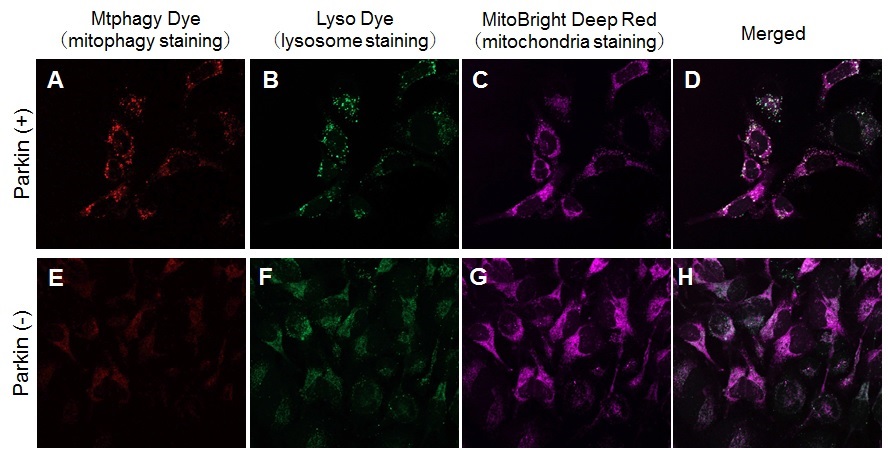
We determined how FK866-induced lysosomal deacidification affects the autophagy-lysosomal pathway. After staining with DAPGreen/DAPRed (for detecting autophagosome), or DALGreen (for detecting autolysosome), HeLa cells were starved in HBSS incubation and then treated with FK866 or Bafilomycin A1. Under the starvation condition, the fluorescent signals from all dyes increased, indicating the proceeding autophagy-lysosomal pathway. On the other hand, only DALGreen's signals were decreased in FK866 and Bafilomycin A1 treated cells with starvation conditions. These results clearly suggested that FK866 inhibits the autophagy-lysosomal pathway by NAD+ depletion-induced lysosomal deacidification.

Nampt inhibitor, FK866 inhibits the progress of autophagosome to autolysosome by lysosomal deacidification. A recent finding shows that the dysfunctional condition of nicotinamide adenine dinucleotide (NAD+) biosynthetic enzyme, Nampt induces lysosomal deacidification1). In this section, we tried to determine how NAD+ depletion-induced lysosomal deacidification affects the autophagy-lysosomal pathway.  1) Mikako Yagi, et. al., EMBO J., 40(8), e105268 (2021)

 Application Note II  (click to open/close)
  Induction of Mitophagy in Parkin Expressed HeLa cells
Induction of mitophagy by carbonyl cyanide m-chlorophenyl hydrazone (CCCP) as a mitochondrial-uncoupling reagent with Parkin expressed HeLa cells.

HeLa cells were seeded on μ-slide 8 well (Ibidi) and cultured at 37oC overnight in a 5%-CO2 incubator. The cells were transfected with Parkin plasmid vector by HilyMax transfection reagent (Code#:H357), and incubated at 37oC overnight. The Parkin expressed HeLa cells were washed with Hanks’ HEPES buffer twice and then incubated at 37oC for 30 minutes with 250 μl of 100 nmol/l Mtphagy Dye working solution containing 100 nmol/l MitoBright LT Deep Red (※). After the washing of the cells with Hanks’ HEPES buffer twice, the culture medium containing 10 μmol/l CCCP was added to the well. After 24 hours incubation, mitophagy was observed by fluorescence microscopy. After removing the supernatant, 250 μl of 1 μmol/l Lyso Dye working solution were added to the cells and incubated at 37oC for 30 minutes. The cells were washed with Hanks’ HEPES buffer twice and then co-localization of Mtphagy, Lyso Dye and MitoBright Deep Red was observed by confocal fluorescence microscopy.

Observation of mitophagy using Parkin
expressed HeLa cells (upper panel) and normal HeLa cells (Lower)
A, E) Fluorescent images of Mtphagy Dye; B, F) Fluorescent images of Lyso Dye;
C, G) Fluorescent images of MitoBright Deep Red (※); D, H) Merged images

※Our sincere apology that we discontinued distributing MitoBright Deep Red.
     Please refer to MitoBright LT Deep Red (Code#:MT12) which improved retention ability.


Previous Science Note  


Product Classification

Product Classification