

|
Recent research on cellular senescence reveals that senescent cells contribute to tumor promotion. Here are some papers that highlight how senescent cells create a pro-tumorigenic microenvironment by enhancing inflammation, angiogenesis, and immune evasion, offering therapeutic opportunities. Cellular senescence is a state of stable cell cycle arrest that prevents the proliferation of damaged or aged cells and acts as a tumor suppressor mechanism. However, senescent cells can secrete a pro-inflammatory and tissue-altering mix of factors that can paradoxically promote cancer progression. Senescent factors contribute to a pro-tumorigenic microenvironment by enhancing inflammation, angiogenesis, and immune evasion. Understanding the dual role of senescence in cancer highlights the complexity of its contribution to tumor suppression and progression and offers potential therapeutic opportunities. |
|||
|
Hematopoietic aging promotes cancer by fueling IL-1⍺–driven emergency myelopoiesis |
Age-dependent loss of HAPLN1 erodes vascular integrity via indirect upregulation of endothelial ICAM1 in melanoma |
Fibroblasts in the Aged Pancreas Drive Pancreatic Cancer Progression Click here for the original article: Daniel J. Zabransky, et. al., Cancer Research, 2024. |
|
|
Point of Interest - DNMT3A (DNA methyltransferase 3A) downregulation with age increases IL-1⍺ production, driving the recruitment of immunosuppressive myeloid cells and tumor progression. - Blocking the IL-1 receptor slows lung cancer progression in aged mice, highlighting potential therapeutic strategies for age-related cancers. |
Point of Interest - ICAM1 leads to VE-cadherin internalization, which increases vascular permeability and melanoma progression. - Blocking ICAM1 reduces tumor size and metastasis in aged mice, highlighting the impact of aging and ECM changes on tumor progression.
|
Point of Interest - Treatment of young mice with GDF-15 enhances tumor growth, whereas GDF-15 knockout in aged mice reduces tumor growth. - AKT inhibition is effective in aged but not young microenvironments, providing a targeted therapy for age-related pancreatic cancer. |
|
| Related Techniques | |||
| Cellular senescence detection | SPiDER-βGal for live-cell imaging or flow cytometry / microplate reader / tissue samples SPiDER-βGal Blue for fixed cell and for multiple staining with immunostaining and other methods |
||
| Total ROS detection | Highly sensitive DCFH-DA or Photo-oxidation Resistant DCFH-DA | ||
| Glycolysis/Oxidative phosphorylation Assay | Glycolysis/OXPHOS Assay Kit | ||
| Oxygen Consumption Rate(OCR) Plate Assa | Extracellular OCR Plate Assay Kit | ||
| First-time autophagy research | Autophagic Flux Assay Kit | ||
| Lysosomal function | Lysosomal Acidic pH Detection Kit-Green/Red and Green/Deep Red | ||
| Apoptosis detection in multiple samples | Annexin V Apoptosis Plate Assay Kit | ||
| Cell proliferation/ cytotoxicity assay | Cell Counting Kit-8 and Cytotoxicity LDH Assay Kit-WST | ||
| Related Applications | |||
Metabolic shift to glycolysis in senescenct cells |
|||
|
|
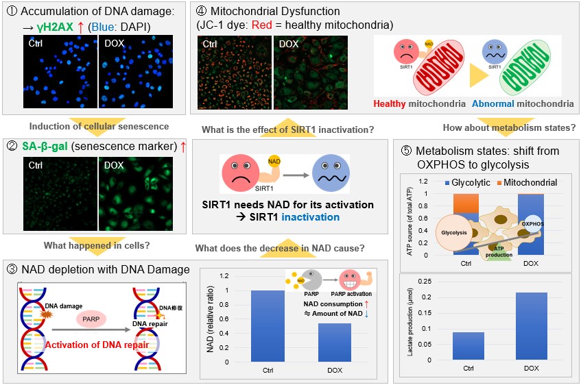
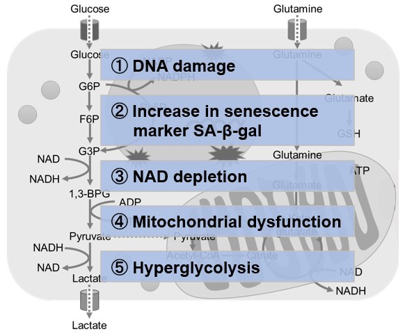
NAD(+) levels decline during the aging process, causing defects in nuclear and mitochondrial functions and resulting in many age-associated pathologies*. Here, we try to redemonstrate this phenomenon in the doxorubicin (DOX)-induced cellular senescence model with a comprehensive analysis of our products. *S. Imai, et al., Trends Cell Biol, 2014, 24, 464-471
|
||
 |
|||
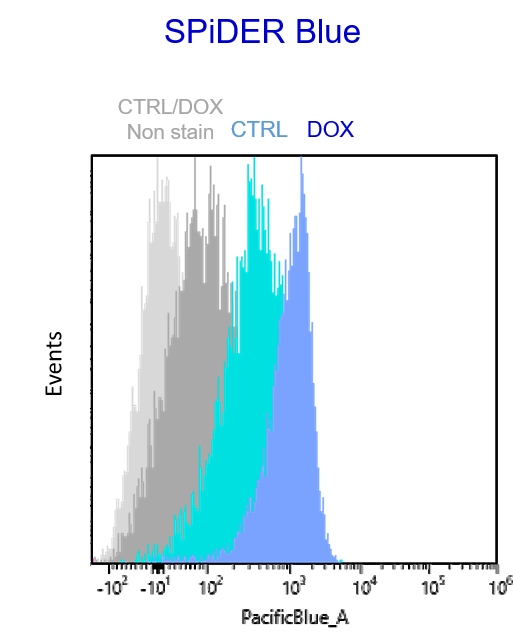
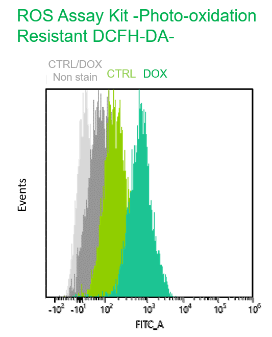
Multiple staining with oxidative stress-related markers using Doxorubicin-induced senescent cells(flow cytometry) |
|||
|
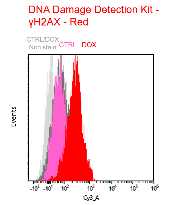
Using A549 cells induced to senescence by doxorubicin (DOX) and normal cells (CTRL), changes in oxidative stress-related markers in senescent cells were analyzed by flow cytometry with multiple staining. SA-βGal as a senescence marker was detected by Cellular Senescence Detection Kit - SPiDER Blue, total ROS as an oxidative stress marker was detected by ROS Assay Kit - Photo-oxidation Resistant DCFH-DA-, and γH2AX as a DNA damage marker was detected by DNA Damage Detection Kit - γH2AX-Red. As a result, total ROS and γH2AX were increased in SA-βGal-positive senescent cells, and the increase in oxidative stress-related markers associated with cellular senescence could be detected by multiple staining. <Experimental Procedure> |
|||


Product Classification
Product Classification
-
Cell Proliferation / Cell Cytotoxicity Assay
Cell Proliferation / Cell Cytotoxicity Assay Kits /Related Reagents
-
Cell Staining
Cell Double Staning Kit /Live Cell Staining /Dead Cell Staining /Nuclear Staining /Mitochondria Staning /Tissue Staining /Nucleolus Staining /Lipid Droplet Staining /Cell Membrane Staining /Lysosome Staining
-
Intracellular Fluorescent Probes
Reagents for Intracellular Calucuum Ion /Reagents for Intracellular Ion /Related Reagents
-
Labeling Chemistry
Protein Labeling Kits /Protein Labeling Reagents /HPLC Derivertization Reagents /Biotion Labeling Reagents /Related Reagents /Exsosome Labeling
-
Oxidative Stress
Stress Maker Detection /NO Detection /NO Donor /NO Inhibitor /ACE Inhibition Assay /Reagents・Kits for Sulfur Biology /Antioxidant Assay Kit /Donors for Sulfur Biology
-
-Bacstain- Series
Bacterial Proliferation Assay Kit /Bacteria Staining /Bacterial Fluorescent Staining
-
Molecular Biology
Transfection Reagents /Nuclear Staining /Agarose /Related Reagents /Buffer for Molecular Biology
-
Detergents
Detergents /Sets
-
Cross-Linking Reagents
Hetero-bifunctional Reagents /Homo-bifunctional Reagents /Others
-
Redox Dyes
Reductive Chromogenic Dyes /Electron Mediators /Oxidative Chromogenic Dyes /Trinder Reagents
-
Ion Analysis
Ionophores /Anion Eliminator /Solvent for Ion Electrode of Liquid Film Type
-
Organic Scintillator
-
Buffers
Buffers
-
Metal Chelates
EDTA /Other Chelator /Reagents for Chelator Titration
-
Chromogen/Metal Indicator
Chromogen/Metal Indicator
-
Water Analysis
/Fluorine /Iron / /Water Hardness /Residual Chlorine /ABS /Cyan / /Chromium /Copper
-
Extraction Reagent
AA Chelator /Related Reagents
-
High Purity Solvent
Spectrozole /Luminazole / /Acnazole /Dehydration Solvent for Synthesis
-
Biochemicals
Biochemicals
-
Functional Organic Material
Alkanethiol Derivative /Phosphonic Acid Derivatives
