Endosomal Proteins Regulating Cellular Fate [Sep. 3, 2024]

[Sep. 3, 2024] Previous Science Note

Endosomal proteins play a critical role in determining cell fate by regulating the trafficking and signaling of receptors and other membrane-bound molecules within cells. These proteins control the endocytosis, recycling, or degradation of signaling receptors, thereby influencing key pathways such as those governing differentiation, survival, and apoptosis. By modulating the duration and intensity of signaling cascades, endosomal proteins help determine whether a cell will proliferate, differentiate, or undergo programmed cell death. Dysregulation of these processes can lead to inappropriate cell fate decisions and contribute to diseases such as cancer and neurodegeneration.

Ku70 senses cytosolic DNA and assembles a tumor-suppressive signalosome
Click here for the original article: Abhimanu Pandey, et. al., Science Advances, 2024.

Point of Interest

- Ku70 deficiency increases susceptibility to colon cancer by disrupting DNA repair and activating aberrant signaling pathways.

- Ku70 forms a complex with GTPase Ras and kinase Raf at Rab5+Rab7+ early-late endosomes, activating MEK-ERK pathways that reduce cell proliferation and tumorigenesis.

- Targeting this Ku70 signalosome could improve cancer treatment by restoring proper DNA repair and cell cycle regulation.

MYCT1 controls environmental sensing in human haematopoietic stem cells
Click here for the original article: Júlia Aguadé-Gorgorió et. al., Nature, 2024.
*ECGreen (Dojindo) was used for FACS-based endocytosis analysis.

Point of Interest

- MYCT1 is a key regulator of human hematopoietic stem cell (HSC) self-renewal and expansion by modulating endocytosis and environmental sensing.

- MYCT1 expression is essential for HSC expansion and engraftment, and its loss leads to excessive endocytosis and dysregulated signaling.

- Restoring MYCT1 in cultured HSCs preserves stemness and improves expansion, highlighting its importance in human HSC regulation.

EGFR-dependent endocytosis of Wnt9a and Fzd9b promotes β-catenin signaling during hematopoietic stem cell development in zebrafish
Click here for the original article: Nicole Nguyen et. al., Science Signaling, 2024.

Point of Interest

- EGFR and EPS15-dependent Wnt9a-Fzd9b signaling is essential for hematopoietic stem cell development in zebrafish.

- EGFR-mediated phosphorylation triggers endocytosis of Wnt9a-Fzd9b complexes, which are necessary for hematopoietic stem cell signaling.

- EPS15 promotes Wnt9a-Fzd9b endocytosis, which is critical for hematopoietic stem cell development in zebrafish and humans.

Related Techniques

Endocytosis Detection detection
ECGreen-Endocytosis Detection
Lysosomal function
Lysosomal Acidic pH Detection Kit -Green/Red and Green/Deep Red
Mitophagy or autophagy detection
Mitophagy Detection KitAutophagic Flux Assay Kit
 
Plasma Membrane Staining
PlasMem Bright Green / Red
Ready-to-use kit for Cell cycle assay
Cell Cycle Assay Solution Blue / Deep Red
Total ROS detection
Highly sensitive DCFH-DA or Photo-oxidation Resistant DCFH-DA

Related Applications

Clear visualization of intracellular vesicular trafficking

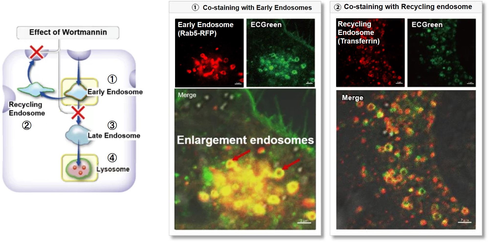
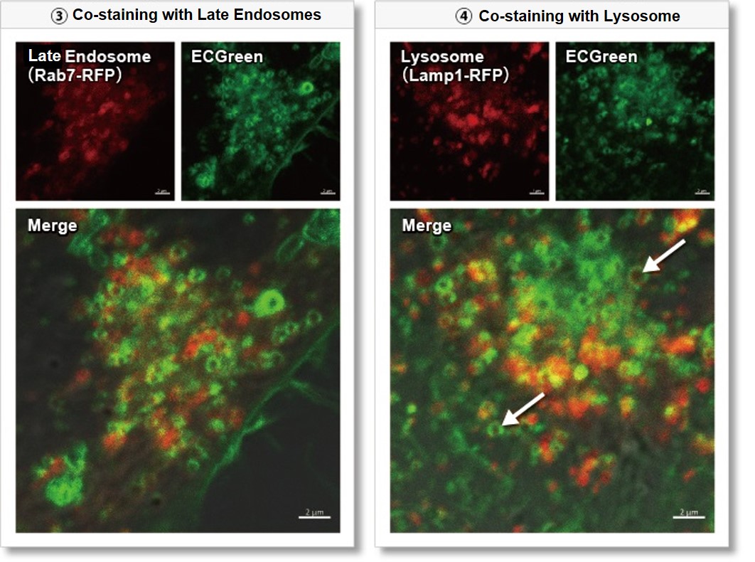
Wortmannin is known to inhibit endosomal recycling and lysosomal translocation, leading to endosomal enlargement.
These changes induced by Wortmannin were confirmed by co-staining with ECGreen (green) and the following indicators.

①Eary endosome: Rab5-RFP (red)
② Recycling endosome: Fluorescent labeled Transferin (red)
③ Late endosome: Rab5-RFP (red)
④ Lysosome: Lamp1-RFP (red)

As a result, it was confirmed that ECGreen (green) co-localizes only with enlarged early endosomes and recycling endosomes (Fig. ① and ②), but not with late endosomes or Lysosomes (Fig. ③ and ④), supporting Wortmannin's effect. ECGreen can visualize changes in the intracellular vesicular trafficking system and endosome shape.

Endosomes (ECGreen, green): Ex. 405 nm / Em. 500 – 560 nm
Early endosomes (Rab5-RFP, red): Ex. 561 nm / Em. 560 – 620 nm
Recycling endosome (Transferrin-Alexa fluor 488 conjugate, red: pseudo-color): Ex. 488 nm / Em. 500 – 550 nm
Late endosomes (Rab7-RFP, red): Ex. 561 nm / Em. 560 – 620 nm
Lysosomes (Lamp1-RFP, red): Ex. 561 nm / Em. 560 – 620 nm
 

[Experimental Procedure]
(1) Prepare HeLa cells in 8 wells of μ-Slide and incubate overnight.
(2) After washing with HBSS, 200 µl of Wortmannin (final concentration: 100 nmol/l) prepared in 10% FBS-containing MEM medium was added.
(3) Incubate at 37°C for 30 minutes
(4) 200 µl of ECGreen (diluted 1,000-fold) prepared in 10% FBS-containing MEM medium without removing the supernatant
(5) Incubate at 37°C for 30 minutes
(6) Wash the cells twice with HBSS and add MEM medium containing 10% FBS.
(7) Observation with a confocal laser microscope

Product Classification

Product Classification