

|
Recent research on cancer progression shows that mtDNA mutations affect cellular metabolism and promote tumor growth. Here are some of the studies showing how mtDNA mutations drive metabolic changes and have implications for cancer therapy. Mitochondrial DNA (mtDNA) plays a key role in cancer progression by influencing cellular metabolism and oxidative phosphorylation. Mutations in mtDNA can lead to metabolic shifts, such as the Warburg effect, that promote tumor growth and alter the tumor microenvironment. These mtDNA alterations can increase the production of reactive oxygen species (ROS), which drive pro-tumor signaling pathways such as TGFβ/Smad. In addition, mtDNA mutations and their transfer via extracellular vesicles (EVs) can affect tumor-stroma communication and potentially serve as biomarkers and therapeutic targets in cancer treatment. |
|
| Related Techniques |
|
|
|
|
|
|
|
|
|
| Related Applications |
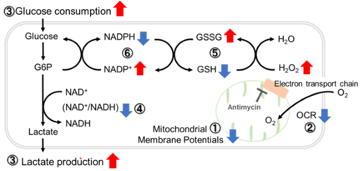
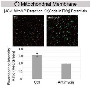
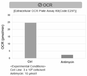
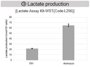
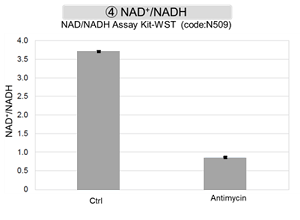
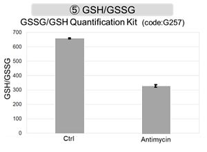
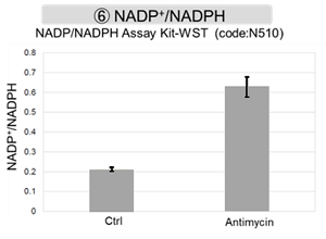
Inhibition of Mitochondrial Electron Transport Chain
|
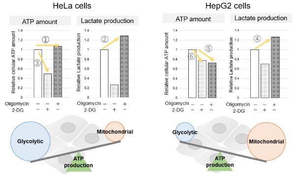
Metabolic Pathway Dependence Across Cell LinesEvaluation by Lactate production and ATP levels
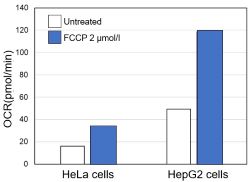
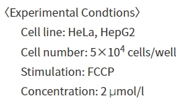
Evaluation by OCR value
|


Product Classification
Product Classification
-
Cell Proliferation / Cell Cytotoxicity Assay
Cell Proliferation / Cell Cytotoxicity Assay Kits /Related Reagents
-
Cell Staining
Cell Double Staning Kit /Live Cell Staining /Dead Cell Staining /Nuclear Staining /Mitochondria Staning /Tissue Staining /Nucleolus Staining /Lipid Droplet Staining /Cell Membrane Staining /Lysosome Staining
-
Intracellular Fluorescent Probes
Reagents for Intracellular Calucuum Ion /Reagents for Intracellular Ion /Related Reagents
-
Labeling Chemistry
Protein Labeling Kits /Protein Labeling Reagents /HPLC Derivertization Reagents /Biotion Labeling Reagents /Related Reagents /Exsosome Labeling
-
Oxidative Stress
Stress Maker Detection /NO Detection /NO Donor /NO Inhibitor /ACE Inhibition Assay /Reagents・Kits for Sulfur Biology /Antioxidant Assay Kit /Donors for Sulfur Biology
-
-Bacstain- Series
Bacterial Proliferation Assay Kit /Bacteria Staining /Bacterial Fluorescent Staining
-
Molecular Biology
Transfection Reagents /Nuclear Staining /Agarose /Related Reagents /Buffer for Molecular Biology
-
Detergents
Detergents /Sets
-
Cross-Linking Reagents
Hetero-bifunctional Reagents /Homo-bifunctional Reagents /Others
-
Redox Dyes
Reductive Chromogenic Dyes /Electron Mediators /Oxidative Chromogenic Dyes /Trinder Reagents
-
Ion Analysis
Ionophores /Anion Eliminator /Solvent for Ion Electrode of Liquid Film Type
-
Organic Scintillator
-
Buffers
Buffers
-
Metal Chelates
EDTA /Other Chelator /Reagents for Chelator Titration
-
Chromogen/Metal Indicator
Chromogen/Metal Indicator
-
Water Analysis
/Fluorine /Iron / /Water Hardness /Residual Chlorine /ABS /Cyan / /Chromium /Copper
-
Extraction Reagent
AA Chelator /Related Reagents
-
High Purity Solvent
Spectrozole /Luminazole / /Acnazole /Dehydration Solvent for Synthesis
-
Biochemicals
Biochemicals
-
Functional Organic Material
Alkanethiol Derivative /Phosphonic Acid Derivatives
