|
Mitochondrial stress responses are a set of cellular reactions triggered when mitochondria, the energy-producing organelles in cells, encounter various forms of stress. This can include damage from reactive oxygen species (ROS), fluctuations in nutrient levels, or other environmental stressors. In response to such stress, mitochondria can undergo changes in their dynamics, such as fission and fusion, to maintain cellular homeostasis and energy production. Additionally, cells activate protective pathways, like the mitochondrial unfolded protein response (UPRmt), to repair or degrade damaged mitochondrial proteins and mitigate the potentially harmful effects of mitochondrial dysfunction.
|
-
Mitochondrial fission drives neuronal metabolic burden to promote stress susceptibility in male mice
Click here for the original article: Wan-Ting Dong, et. al., Nature Metabolism, 2023.
Point of Interest
- Inhibition of Drp1 in neurons ameliorates stress-related depressive-like behavior.
- Enhancing Drp1-dependent mitochondrial fission promotes stress susceptibility.
- Increased stress susceptibility is alleviated by increasing mitochondrial ATP production.
-
Mitochondrial DNA breaks activate an integrated stress response to reestablish homeostasis
Click here for the original article: Yi Fu, et. al., Molecular Cell, 2023.
Point of Interest
- Mitochondrial double-strand breaks (mtDSBs) lead to significant mitochondrial defects and impede protein import.
- mtDSBs initiate an integrated stress response (ISR).
- Inhibition of the ISR exacerbates mitochondrial defects and slows recovery of mitochondrial DNA.
- ATAD3A is a potential signaling molecule from damaged genomes to the inner membrane of mitochondria.
-
Glial-derived mitochondrial signals affect neuronal proteostasis and aging
Click here for the original article: Raz Bar-Ziv et. al., Science Advances, 2023.
Point of Interest
- The mitochondrial unfolded protein response (UPRmt) activation in four astrocyte-like glial cells can alleviate protein aggregation in neurons.
- Glial cells use small clear vesicles (SCVs) to signal the mitochondrial proteotoxic stress to neurons.
- Neurons relay the signal to the periphery using dense-core vesicles.
|
| Related Techniques |
|
|
|
|
|
|
|
|
|
|
|
|
|
|
- Mitochondrial staining (Long-Term Visualization)
- MitoBright LT Green, Red, Deep Red
|
|
|
| Related Applications |
Lysosomal Function and Mitochondrial ROS
-
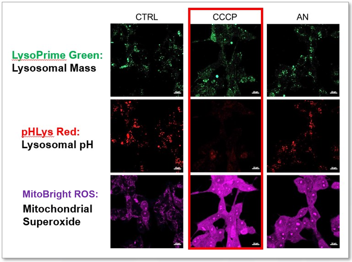
CCCP and Antimycin are recognized inducers of mitochondrial ROS, linked to the loss of mitochondrial membrane potential. Recent studies have shown that CCCP induces not only mitochondrial ROS but also lysosomal dysfunction. To observe mitochondrial ROS, HeLa cells were labeled with MitoBright ROS Deep Red for Mitochondrial Superoxide Detection, and the lysosomal mass and pH were independently detected with LysoPrime Green and pHLys Red. Co-staining with MitoBright ROS and Lysosomal dyes revealed that CCCP, unlike Antimycin, triggers concurrent lysosomal neutralization and mitochondrial ROS induction.
Reference: Benjamin S Padman, et. al., Autophagy (2013)
Products in Use
- LysoPrime Green
- pHLys Red
- Lysosomal Acidic pH Detection Kit
- MitoBright ROS - Mitochondrial Superoxide Detection
|
Lysosomal Function and Mitochondrial ROS
-
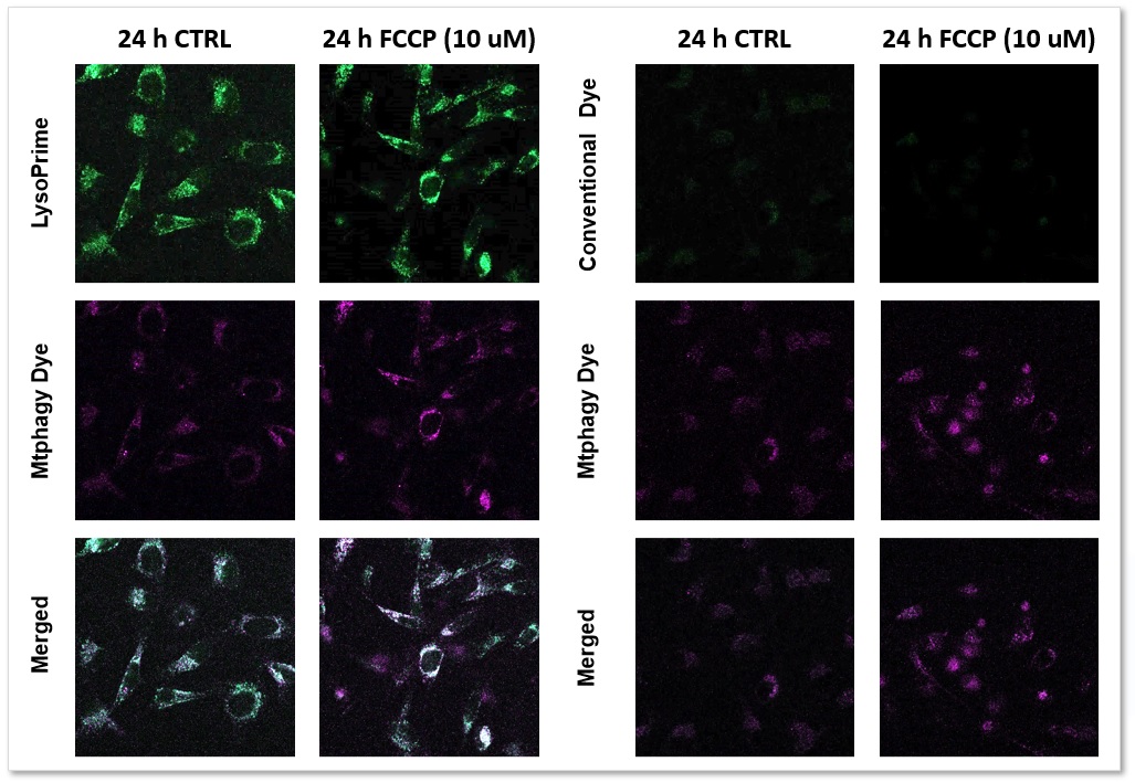
We performed fluorescence imaging by stimulating mitophagy induction in SHSY-5Y cells stained with Mitophagy Detection(Code: MD01) and LysoPrime Green or existing products. The fluorescence signal of LysoPrime Green did not decrease and the lysosomal localization over time was confirmed. This means that the co-localization rate of the fluorescent spots of the Mtphagy Dye is higher than that of the existing product, and thus more accurate mitophagy analysis can be performed.
LysoPrime Green: Ex= 488 nm, Em= 500-570 nm
Mtphagy Dye: Ex= 561 nm, Em= 560-650 nm
Products in Use
- LysoPrime Green
- Mitophagy Detection Kit and Mtphagy Dye
|
Inhibition of Mitochondrial Electron Transport Chain
-
-
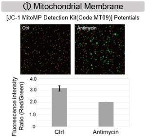
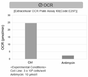
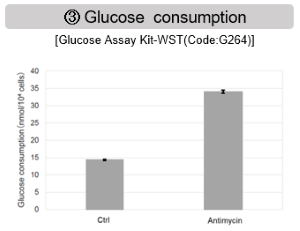
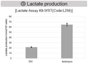
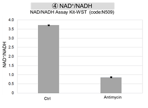
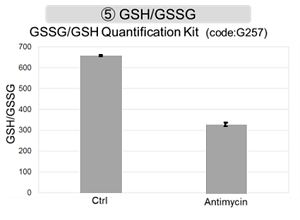
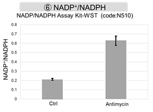
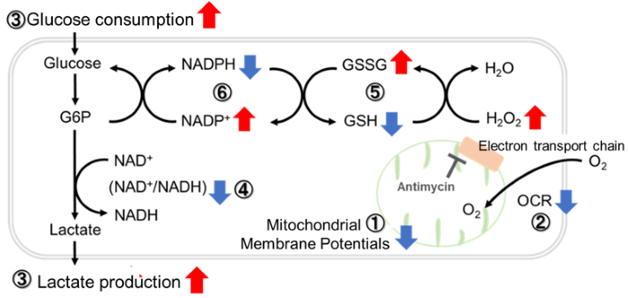
Antimycin stimulation of Jurkat cells was used to evaluate the changes in cellular state upon inhibition of the mitochondrial electron transport chain using a variety of indicators.
The results showed that inhibition of the electron transport chain resulted in (1) a decrease in mitochondrial membrane potential and (2) a decrease in OCR. In addition, (3) the NAD+/NADH ratio of the entire glycolytic pathway decreased due to increased metabolism of pyruvate to lactate to maintain the glycolytic pathway, (4) GSH depletion due to increased reactive oxygen species (ROS), and (6) increase in the NADP+/NADPH ratio due to decreased NADH required for glutathione biosynthesis were observed.
|


