|
Senescence is known to drive iron accumulation and promote ferroptosis. However, recent research on iron dynamics reveals that senescence can also lead to functional iron deficiency, conferring resistance to ferroptosis. Here are some papers that provide insight into the role of iron dynamics and ferroptosis in senescence.
Cellular senescence, iron accumulation, and ferroptosis are intricately linked processes that influence aging and tissue degeneration. Traditionally, senescent cells are known to accumulate iron, particularly ferritin-bound iron in lysosomes, which increases labile iron levels and promotes the production of reactive oxygen species that drive ferroptosis and contribute to tissue damage. However, recent evidence suggests that aging can also lead to functional iron deficiency through pathways such as NUPR1-lipocalin-2, reducing stemness and tumorigenic potential in aged cells while conferring resistance to ferroptosis. These findings highlight the dual role of iron in senescence and ferroptosis and underscore the need for further research to fully understand these complex mechanisms.
|
-
Ageing limits stemness and tumorigenesis by reprogramming iron homeostasis
Click here for the original article: Xueqian Zhuang, et. al., Nature, 2024.
Point of Interest
- Aging induces the transcription factor NUPR1-lipocalin-2, leading to iron deficiency, loss of stemness and reduced tumorigenic potential in aged alveolar cells.
-Inactivation of NUPR1-lipocalin-2 or iron supplementation restores stemness and promotes tumorigenic potential in aged cells, but induces ferroptosis in young alveolar cells.
-These findings highlight the role of age-associated iron deficiency in cancer resistance and the need for youth-focused cancer prevention strategies.
-
Iron accumulation drives fibrosis, senescence and the senescence-associated secretory phenotype
Click here for the original article: Mate Maus et. al., Nature Metabolism, 2023.
Point of Interest
- Senescent cells accumulate iron and drive fibrosis via their secretome, known as the senescence-associated secretory phenotype (SASP), and reactive oxygen species, even when extracellular iron levels are normal.
- Vascular and hemolytic injury induces iron accumulation and promotes cellular senescence and fibrosis in mice and humans.
- Iron metabolism is a therapeutic target for senescence-related diseases, and iron detection provides a non-invasive assessment of fibrosis.
-
Aging promotes metabolic dysfunction-associated steatotic liver disease by inducing ferroptotic stress
Click here for the original article: Kuo Du et. al., Nature Aging, 2024.
Point of Interest
- A gene cluster characteristic of aging hepatocytes is enriched in diseased livers and includes genes that induce ferroptosis.
- Aged mice show increased hepatocyte ferroptosis and liver degeneration under conditions that induce metabolic stress.
- Inhibition of ferroptosis in aged mice reverses age-related liver damage.
|
|
Related Techniques
|
- Intracellular / mitochondrial ferrous ion (Fe2+) detection
- FerroOrange(intracellular), Mito-FerroGreen(mitochondrial)
|
- Cellular senescence detection
- SPiDER-βGal for live-cell imaging or flow cytometry / microplate reader / tissue samples
SPiDER-βGal Blue for fixed cell and for multiple staining with immunostaining and other methods
|
|
|
- Lipid Droplet detection
- Lipid Droplet Assay Kit - Blue / Deep Red
|
- Total ROS detection
- Highly sensitive DCFH-DA or Photo-oxidation Resistant DCFH-DA
|
- Mitochondrial superoxide detection
- MitoBright ROS Deep Red - Mitochondrial Superoxide Detection
|
- Glutathione Quantification
- GSSG/GSH Quantification Kit
|
- Cystine Uptake detection
- Cystine Uptake Assay Kit
|
- MDA detection
- MDA Assay Kit
|
- Glycolysis/Oxidative phosphorylation Assay
- Glycolysis/OXPHOS Assay Kit
|
- Apoptosis detection in multiple samples
- Annexin V Apoptosis Plate Assay Kit
|
- Cell proliferation/ cytotoxicity assay
- Cell Counting Kit-8 and Cytotoxicity LDH Assay Kit-WST
|
|
Related Applications
|
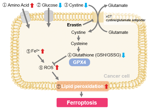
Erastin-Induced Ferroptosis: Evaluating Intracellular Uptake and Redox Balance
We investigated the transition of cellular metabolisms in A549 cells treated with erastin, a known ferroptosis inducer. Our results revealed the following.

- The inhibition of cystine uptake by erastin led to a depletion of cysteine, which in turn increased the compensatory uptake of other amino acids.
- Glucose uptake, which typically promotes ferroptosis*, was found to decrease upon erastin treatment, suggesting a potential cellular self-defense mechanism.
- The depletion of cysteine resulted in a decrease in glutathione levels and an increase in Fe2+, ROS, and lipid peroxides, all of which are recognized markers of ferroptosis.
Cell Line: A549
Incubation Conditions: 100 μmol/l Erastin/MEM, 37℃, 3h
*Reference: Xinxin Song, et al., Cell Reports, (2021)
Products in Use
|


