| [July. 5, 2023] | Previous Science Note |
|
Scientists have unveiled that in comparison to young mice, one-third of old microglia show Lipofustin-related autofluorescence (AF), characterized by profound changes in lipid and iron content, phagocytic activity, and oxidative stress. Pharmacological removal of microglia in older mice successfully eliminated AF-microglia, following the repopulation of new functional microglia leads to an improvement in age-related neurological impairments and reduces neurodegeneration after traumatic brain injury. Learn more about how the authors phenotyped AF-microglia using Lipi-Blue* for Lipid droplet labeling, and FerroOrange* for iron labeling. (Please refer to Fig. 1E, 5F, 9E for FerrOrange, Fig. 5D, 7E, for Lipi-Blue) |
|
|
Related Techniques |
|
|
|
|
|
|
|
Related Applications |
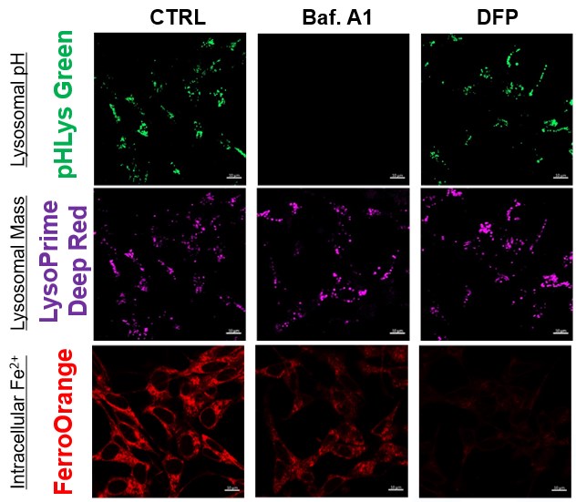
The simultaneous detection of lysosomal function with intracellular Fe2+
Recent reports suggest that lysosomal neutralization can result in iron depletion, consequently leading to the disruption of cell viability. To verify this, HeLa cells were labeled with FerroOrange for Fe2+ detection, and the lysosomal mass and pH were separately detected with LysoPrime DeepRed and pHLys Green (a product currently under development). Co-staining with FerroOrange and Lysosomal dyes demonstrated that Bafilomycin A1 (Baf. A1), an inhibitor of lysosomal acidification, causes iron depletion consistent with the findings reported in the article. Interestingly, the iron chelator, Deferiprone (DFP), did not impact lysosomal pH, suggesting that lysosomal function plays a key role in managing iron homeostasis. Reference: Ross A Weber, et. al., Mol Cell (2020) Products in Use *pHLys Green is available as the "Lysosomal Acidic pH Detection Kit-Green/Deep Red". |