


-SulfoBiotics- Biotin-HPDP(WS) solution
Biosulfur Analysis
-
Product codeSB17 -SulfoBiotics- Biotin-HPDP(WS) solution
-
CAS No.-
| Unit size | Price | Item Code |
|---|---|---|
| 500 µl | $278.00 | SB17-10 |
Description
Biotin-HPDP(WS) is a novel water-soluble biotin-labeling reagent that is modified from Biotin-HPDP (Product code: B573). Biotin-HPDP is a conventional reagent to introduce a biotin moiety to a sulfhydryl group of protein via reversible disulfide bond. This reagent is useful for affinity-purification of biotinylated proteins by using avidin-coated beads, because the disulfide bond is cleavable by a reducing agent (Fig.1). Thus, Biotin-HPDP has been widely used for biotin switch assay, which is an analytical technique for protein thiol modifications such as s-nitrosylation, s-sulfhydration, and s-palmitoylation. However, preparation of the solution is time-consuming due to the low-solubility in water, even DMSO and DMF. Therefore, Biotin-HPDP(WS) solution was developed to improve the solubility of Biotin-HPDP.
-SulfoBiotics- Biotin-HPDP(WS) solution is an easy-to-use aqueous solution including 20mmol/l Biotin-HPDP(WS).

Figure 1 Schematic Protocol for Biotin Labeling with Biotin-HPDP(WS) and Purifiation of the Biotinylated Protein
| Developer | Dojindo Molecular Technologies, Inc. |
|---|

Manual
Technical info
< Procedure >
1. Lysis buffer (35 μl) and 100 mmol/l DTT in Lysis buffer (5μl) were added to 10 μl of 1 mg/ml GAPDH solution in PBS in a 1.5 ml-microtube, and mixed by vortex.
2. After the solution was incubated at 37 ℃ for 30 minutes, the whole solution was transferred to a 10 K filtration tube, and centrifuged at 12,000 rpm for 10 minutes.
3. PBS (50 μl) was added to the filtration tube, and centrifuged at 12,000 rpm for 10 minutes.
4. Step 3 was repeated.
5. RIPA buffer (126 μl) and 4 mmol/l Biotin-HPDP(WS) solution in H2O (14 μl) were added to the filtration tube, and mixed with the protein by pipetting.
6. The filtration tube was incubated at 37 ℃ for 1 hour, and centrifuged at 12,000 rpm for 10 minutes.
7. PBS (50 μl) was added to the tube, and centrifuged at 12,000 rpm for 10 minutes.
8. Step 7 was repeated.
9. Neutralization buffer (400 μl) was added to the tube to dissolve the biotin-labeled protein by pipetting, and the solution was transferred to a 1.5 ml-microtube.
10. The solution (50 μl) of Step 9 was added to NeutravidinTM Agarose beads in a tube. ※ Neutravidin Agarose beads were washed with Neutralization buffer prior to the reaction.
11. The solution was incubated at 4 ℃ for 1 hour.
12. The tube was centrifuged at 2,500 rpm for 1 minute, and the supernatant was removed using a pipette.
13. Neutralization buffer (+600 mmol/l NaCl) (1 ml) was added to the tube and centrifuged at 2,500 rpm for 1 minute, andthe supernatant was removed using a pipette.
14. Step 13 was repeated twice.
15. Neutralization buffer (1 ml) was added to the tube and centrifuged at 2,500 rpm for 1 minute, and the supernatant was removed using a pipette.
16. Step 15 was repeated.
17. Elution buffer (50 μl) was added to the tube, and mixed by vortex. The solution was incubated at 4 ℃ for 1 hour.
18. The tube was centrifuged at 2,500 rpm for 1 minute, and 10 μl of the supenatant was transferred to a 1.5 ml-microtube.
19. Loading buffer (2 μl) was added to the microtube, and the solution was applied to SDS-PAGE (CBB staining) and western blotting.
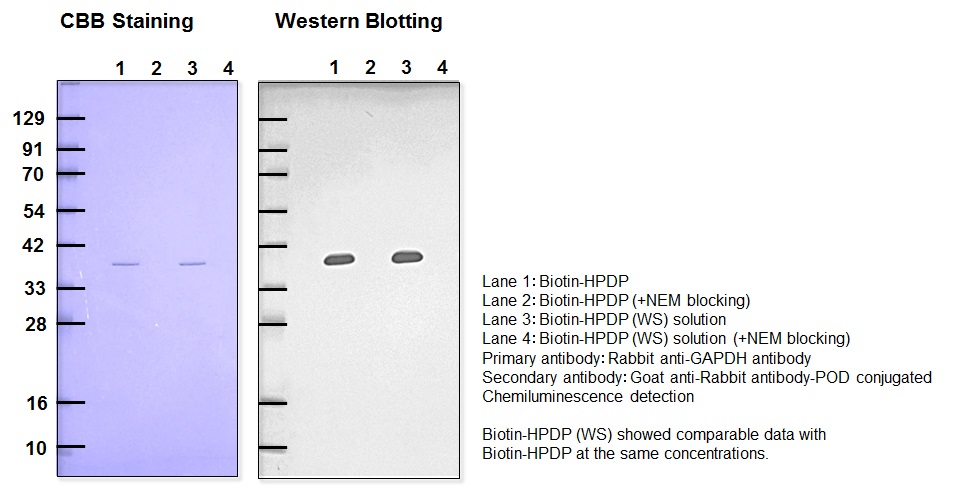
Figure 2 Detection of recovered GAPDH
Comparison between Biotin-HPDP and Biotin-HPDP (WS)
References
1) S. R. Jaffrey and Solomon H. Snyder, "The biotin switch method for the detection of S-nitrosylated proteins ", Sci. STKE, 2001, 86, pl1.
2) X. Wang, N. Kettenhofen, S. Shiva, N. Hogg, and M. Gladwin, "Copper dependence of the biotin switch assay: modified assay for measuring cellular and blood nitrosated proteins", Free Radic. Biol. Med., 2008, 44, 1362.
3) M. T. Forrester, M. W. Foster, M. Benhar, and J. S. Stamler, "Detection of protein S-nitrosylation with the biotin switch technique", Free Radic. Biol. Med., 2009, 46(2), 119.
4) M. D. Kornberg, N. Sen, M. R. Hara, K. R. Juluri, J. V. K. Nguyen, A. M. Snowman, L. Law, L. D. Hester, and S. H. Snyder, "GAPDH mediates nitrosylation of nuclear proteins", Nat. Cell Biol., 2010, 12(11), 1094.
5) J. Wan, A. F. Roth, A. O. Bailey, and N. G. Davis, "Palmitoylated proteins: purification and identification", Nat. Protoc., 2007, 1573.
6) A. K. Mustafa, M. M. Gadalla, N. Sen, S. Kim, W. Mu, S. K. Gazi, R. K. Barrow, G. Yang, R. Wang, and S. H. Snyder, "H2S signals through protein S-sulfhydration", Sci. Signal., 2009, 2(96), ra72.
Handling and storage condition
| Appearance: | Colorless to slightly yellow liquid |
|---|---|
| Dye content: | 0.40 - 0.44 (around 283 nm) |
| 0-5°C |


