


IntactMito Fractionation Kit for Tissue

IntactMito Fractionation Kit for Tissue
- Mitochondria can be fractionated from excised organs within approximately 2 hours
- Enables isolation of highly active mitochondria (OCR, i.e., Oxygen Consumption Rate, measurable)
- Includes three dedicated buffers and centrifuge tubes for the procedure
-
Product codeMT17 IntactMito Fractionation Kit for Tissue
| Unit size | Price | Item Code |
|---|---|---|
| 10 tests | $380.00 | MT17-10 |
[Notice]
To evaluate mitochondrial activity, it is necessary to add succinate or other substrates. For details, please refer to the technical information on this page or the FAQ section under “Are there any precautions when evaluating fractionated mitochondria?”.
| 10 tests | ・Homogenization Buffer ・Resuspension Buffer ・Stabilization Buffer ・Reagent A ・Reagent B ・Centrifuge Tube |
50 ml ×1 15 ml ×1 15 ml ×1 150 g ×2 5 g ×1 30 tubes |
|---|


Activity Evaluation Using Intact Mitochondria Derived from Tissues
To gain a deeper understanding of pathological conditions, it is important not only to assess mitochondrial activity at the cellular level, but also to evaluate mitochondrial activity within tissues.
This kit enables fractionation of mitochondria from tissues while maintaining their activity (i.e., in an intact state). By analyzing these fractionated mitochondria, you can directly evaluate mitochondrial activity which reflects aging, pathological conditions, and even changes induced by drug treatments.
Products Related to Mitochondrial Activity Evaluation
| Product Name | Target | Detection Properties |
|---|---|---|
| Extracellular OCR Plate Assay Kit | Extracellular Oxygen Concentration | Plate Reader Ex: 500 nm / Em: 650 nm |
| JC-1 MitoMP Detection Kit | Mitochondrial Membrane Potential | Fluorescence Microscope, FCM, Plate Reader Green:Ex 488 nm / Em 500-550 nm Red:Ex 561 nm / Em 560-610 nm |
| ATP Assay Kit-Luminescence | ATP | Plate Reader Luminescence |
| MitoComplex-I Activity Assay Kit | Complex I Activity | Plate Reader Colorimetric, λ= 340 nm |

Manual
Technical info
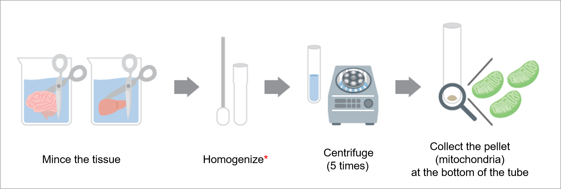
Mitochondrial Fractionation Procedure
This kit includes three dedicated buffers and centrifuge tubes, enabling the fractionation of intact mitochondria from excised organs in approximately two hours.
Please note: A homogenizer* is not included in this kit.
This product was commercialized under the guidance of Dr. Shigeomi Shimizu and Dr. Akira Torii, who are affiliated with Institute of Science Tokyo.
Fractionation of Intact Mitochondria
Using this kit, we added succinate to mitochondria fractionated from mouse brain and performed OCR (oxygen consumption rate) measurements.
The results confirmed that this kit enables fractionation of intact mitochondria that maintain oxygen consumption capacity.
OCR Measurement of Mitochondria Fractionated from Mouse Brain Tissue
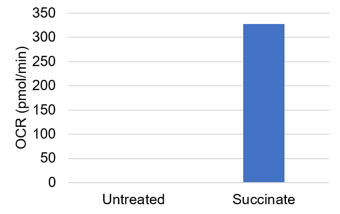
<Experimental Conditions>
Amount of mitochondria: 50 μg/well (as protein levels)
Succinate: 10 mmol/l
Product used: Extracellular OCR Plate Assay Kit (Product Code: E297)
Fractionation of More Intact Mitochondria Compared with a Conventional Kit
Mitochondria were fractionated from mouse brain tissue using a conventional kit and Dojindo’s product, and oxygen consumption was measured adding succinate as a substrate. In addition, ADP and FCCP were applied to stimulate mitochondrial respiration, and the basal and maximal oxygen consumption of the fractionated mitochondria were evaluated and compared.
As a result, mitochondria fractionated using Dojindo’s product showed a clear difference between basal and maximal oxygen consumption, suggesting that a greater proportion of intact mitochondria was obtained.
| Dojindo Laboratories(MT17) | Campany T(Conventional Kit) | |
|---|---|---|
|
|
 |
|
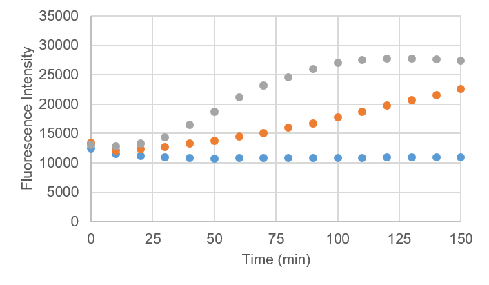

<Experimental Conditions>
Amount of mitochondria: 50 μg/well (as protein levels)
Product used: Extracellular OCR Plate Assay Kit (Product Code: E297)
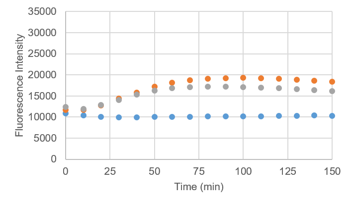
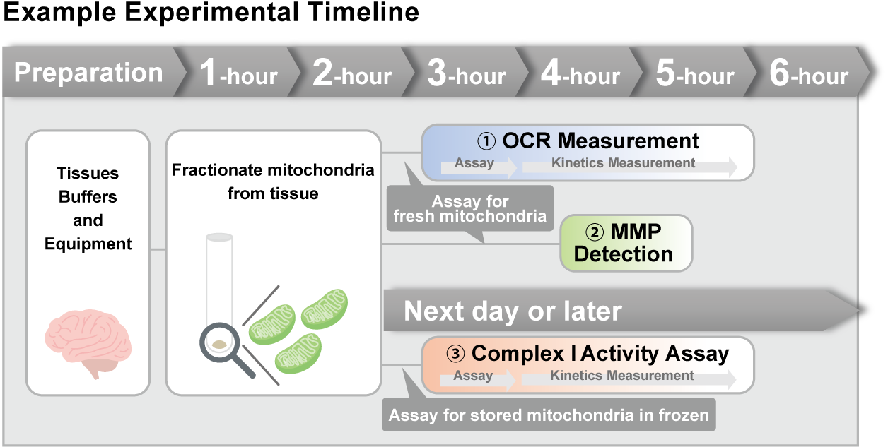
Example Experiment: Activity Evaluation of Mitochondria Fractionated from Mouse Brain
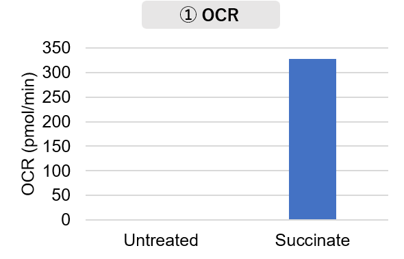
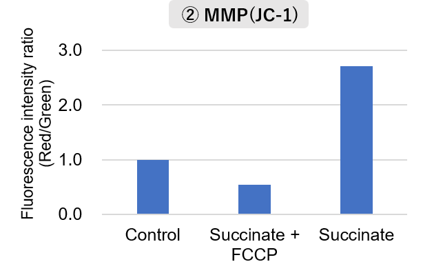
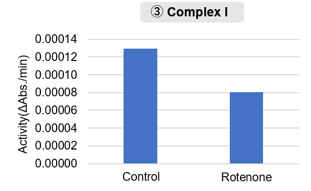
Mitochondria were fractionated from mouse brain tissue using this kit, and oxygen consumption rate (OCR), mitochondrial membrane potential (MMP), and Complex I activity were measured.
The results showed that the addition of succinate, a substrate that activates Complex II of the electron transport chain, increased both OCR and MMP. In contrast, FCCP treatment reduced MMP, indicating that intact mitochondria were successfully fractionated.
Furthermore, in the Complex I activity assay, a decrease in activity was observed following treatment with rotenone, a Complex I inhibitor.
<Experimental Conditions>
OCR Measurement
Amount of mitochondria: 50 μg/well (as protein levels)
Succinate: 10 mmol/l
MMP Detection
Amount of mitochondria: 50 μg/well (as protein levels)
Succinate: 10 mmol/l, FCCP: 4 μmol/l
Complex I Activity Assay
Amount of mitochondria: 20 μg/well (as protein levels)
Rotenone: 10 μmol/l
<Product used>
Mitochondrial Fractionation: IntactMito Fractionation Kit for Tissue (Code: MT17)
OCR measurement: Extracellular OCR Plate Assay Kit (Code: E297)
MMP detection: JC-1 MitoMP Detection Kit (Code: MT09)
Complex I activity assay: MitoComplex- I Activity Assay Kit (Code: MT18)
Q & A
-
Q
Is it possible to fractionate mitochondria from cells?
-
A
No. Since the kit protocol and centrifuge tubes are not compatible with cultured cell samples, mitochondria cannot be fractionated from cells.
-
Q
Which organs have been used with this kit?
-
A
Mitochondria have been successfully fractionated from brain, liver, and heart tissues using this kit.
Please refer to the user manual for fractionation procedures from brain and liver.
Fractionation from heart tissue requires additional preprocessing. If you are interested in this option, please contact our customer support team.
-
Q
How much tissue is required, and how many mitochondria can be fractionated from it?
-
A
Based on our internal experimental results, the mitochondrial yield obtained from mouse brain and liver tissues is as follows:
Brain: Approximately 5 mg of mitochondria (by protein content) were obtained from about 0.4 g of tissue.
Liver: Approximately 7 mg of mitochondria (by protein content) were obtained from about 0.8 g of tissue.Additionally, when 1.2 g of brain tissue (equivalent to three mice) was used, around 10 mg of mitochondria (by protein content) was obtained. Please note that increasing the amount of tissue does not necessarily result in a proportional increase in mitochondrial yield.
-
Q
Are there any differences in the fractionated mitochondria between different organs?
-
A
Yes. Mitochondria fractionated using this kit may exhibit organ-specific variations.
Using brain and liver tissues, we examined the levels of nuclear, cytosolic and mitochondrial markers at each of the four fractionation steps using Western blotting.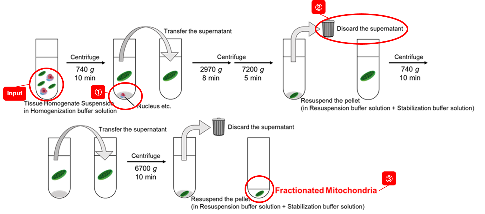
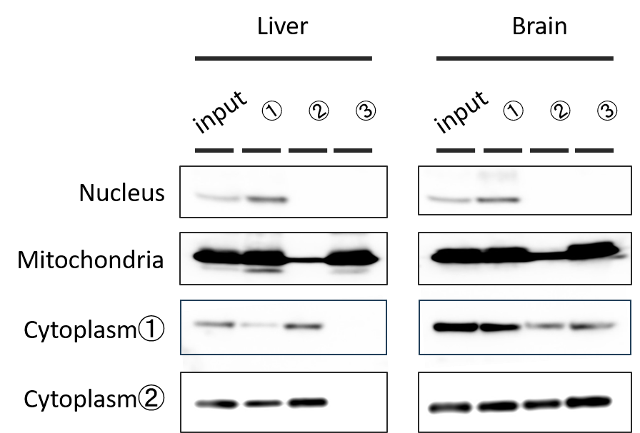
<Protein Loaded>
10 µg/lane<Markers Used>
Nuclear: Lamin A/C (60–80 kDa)
Mitochondrial: COX IV (10–20 kDa)
Cytosolic ①: β-Actin (40–50 kDa)
Cytosolic ②: GAPDH (30–40 kDa)In the final fraction (③), mitochondrial markers were detected in both brain and liver samples, confirming successful mitochondrial fractionation.
However, in brain samples, cytosolic components were also present in the final fraction.
We recommend checking the purity of the final mitochondrial fraction in advance when comparing mitochondrial activities between different organs.
-
Q
Are there any precautions when evaluating fractionated mitochondria?
-
A
For structural evaluations of mitochondria—such as enzyme activities of respiratory complexes or protein quantification—the fractionated mitochondria can be used directly.In contrast, functional evaluations (e.g., oxygen consumption rate or membrane potential) require the addition of appropriate substrates.
We have confirmed successful measurements of oxygen consumption rate and mitochondrial membrane potential by adding succinate.Preparation of Succinate Solution
- A succinate stock solution (0.25 mol/l aqueous succinate, neutralized with 5 mol/l KOH) was diluted with the Stabilization Buffer solution to the desired working concentration.
-
Q
How can I quantify the sample (fractionated mitochondria)?
-
A
We have experience quantifying protein concentration using the BCA assay.
Because the buffers used for fractionation contain proteins to stabilize mitochondria, each buffer includes an equivalent amount of protein. Therefore, when performing protein quantification, please measure the blank using the Stabilization Buffer solution alone, and subtract this value from your sample readings to obtain the correct protein concentration.
Handling and storage condition
| 0-5°C, Protect from moisture |


