 Hidden sections will not be printed.
General Information
It has been recognized that hydrogen sulfide (H2S) has an important role as a physiological active substance for vasodilation, cytoprotection, and modulation of insulin secretion. H2S is considered as a gaseous molecule such as nitric oxide and carbon monoxide. However, around 80% of the total sulfide exists as hydrogen sulfide anion (HS-) under physiological condition, since the pKa is about 7. In addition, HS- easily converts to various biochemical molecules such as persulfides and polysulfides, which react with sulfhydryl moieties in a living body. -SulfoBiotics- HSip-1 DA is cell membrane permeable and it enables fluorescent imaging of intracellular H2S.
-
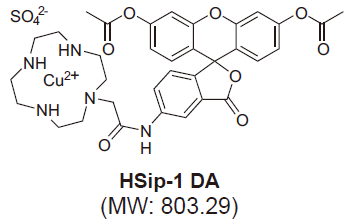
Fig. 1 Chemical structures of HSip-1 DA
-
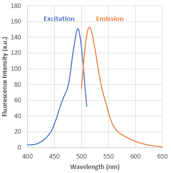
λex : 491 nm
λem : 516 nm
<Recommended filter>
Ex:470 ~ 500 nm
Em:500 ~ 550 nmFig. 2 Excitation and emission spectra of HSip-1 reacted with H2S
Contents
| -SulfoBiotics- HSip-1 DA | 50 μg x 1 |
Storage Conditions
Store at -20oC.
Required Equipment and Materials
- Dimethyl sulfoxide (DMSO)
- Serum-free medium
- HBSS
- Micropipettes
Preparation of Solutions
Preparation of 1 mmol/l HSip-1 DA stock solution
Add 62 μl of DMSO to a tube containing 50 μg of HSip-1 DA and dissolve it by pipetting.
- Store at -20 °C. The reconstituted solution is stable at -20oC for 1 month.
Experimental Example
Fluorescence imaging of hydrogen sulfide with HSip-1 DA
- HeLa cells were seeded on μ-slide 8 well (Ibidi) and cultured at 37oC overnight in a 5% CO2 incubator.
- The culture medium was discarded and the cells were washed with a serum-free medium (MEM) twice.
- HSip-1 DA stock solution (1 mmol/l) was diluted with a serum-free medium (MEM) to prepare 5 μmol/l HSip-1 DA working solution.
- Please optimize the final concentration of HSip-1 DA depeneding on the cell lines.
- HSip-1 DA working solution (5 μmol/l, 200 μl) was added to the cells, and the cells were cultured at 37oC for 30 minutes in a 5% CO2 incubator.
- The supernatant was discarded, and the cells were washed with HBSS twice.
- Na2S solution (200 μmol/l, 200 μl) was added to the each well, and the cells were cultured at 37oC for 30 minutes in a 5% CO2 incubator.
- The supernatant was discarded and the cells were washed with HBSS twice.
- HBSS (200 μl) were added, and the cells were observed by confocal fluorescence microscopy.
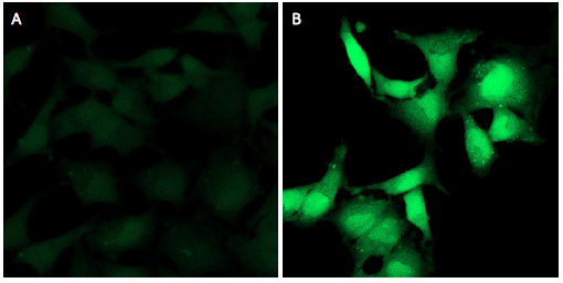
Fig.3 Detection of hydrogen sulfide using HSip-1 DA in HeLa cells treated with Na2S.
(A: Control, B: 200 μmol/l Na2S treated)
These products were commercialized under the advisory of Dr. Tetsuo Nagano and Dr. Kenjiro Hanaoka (The University of Tokyo).
Reference
- K. Sasakura, K. Hanaoka, N. Shibuya, Y. Mikami, Y. Kimura, T. Komatsu, T. Ueno, T. Terai, H. Kimura, and T. Nagano, “Development of a Highly Selective Fluorescence Probe for Hydrogen Sulfide”, J. Am. Chem. Soc., 2011, 133, 18003.
Frequently Asked Questions / Reference


SB22: -SulfoBiotics- HSip-1 DA
Revised Aug., 10, 2023