 Hidden sections will not be printed.
General Information
The plasma membrane (PM), consists of a lipid bilayer separating the intracellular environment from the extracellular space. Consequently, the PM plays a central role in many cell behaviors, such as cell migration, cell stretching, and signaling cascades. Additionally, PM dysfunction is an important biomarker because it is related to the cell status and is linked to many diseases1).
Methods for monitoring PM morphology and dynamics are usually based on labeling the PM with small fluorescent molecules, expressing fluorescent protein targeted to the PM via plasmid transfection, or immunostaining the PM with an antibody. Because the latter two methods have major limitations, such as the requirement for stable expression of the fluorescent protein and their applicability for fixed cells only, small fluorescent molecule dyes, which can simply be added to live cells, are widely used2). However, these dyes also have some limitations, including the short duration of their retention in the PM and the requirement of a special solution with which to dilute the dyes because of their low water solubility3).
Dojindo’s PlasMem Bright dyes overcome these limitations. PlasMem Bright dyes are designed to stain PMs for over a day. Furthermore, the PlasMem Bright dyes are more water-soluble compared with other commercially available dyes and can be diluted with culture medium. The PlasMem Bright dyes offer two different color options (green and red) and are provided as ready-to-use DMSO solutions. A working solution can be prepared easily via a single dilution step using growth medium or HBSS.
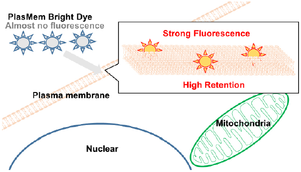
Figure 1. Mechanism of cell staining by PlasMem Bright dyes.
Fluorescent Properties
Excitation and emission spectra of PlasMem Bright dyes
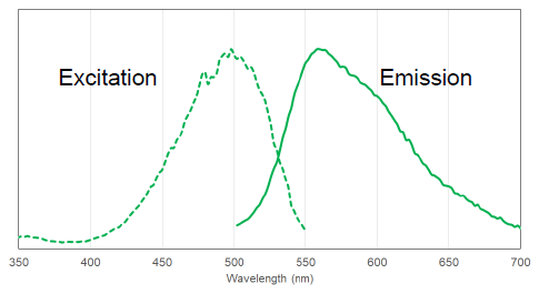
Figure 2. Excitation and Emission spectra of PlasMem Bright Green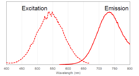
Figure 3. Excitation and Emission spectra of PlasMem Bright Red
Contents
| P504 PlasMem Bright Green | 100 µl x 1 |
| P505 PlasMem Bright Red | 100 µl x 1 |
Storage Condition
| P504 PlasMem Bright Green | Store at -20 oC, protect from light and moisture. |
| P505 PlasMem Bright Red | Store at -20 oC, protect from light and moisture. |
Required Equipment and Materials
- Growth medium or HBSS
- Micropipettes
- Microtube
Preparation of Solutions
Preparation of PlasMem Bright working solution
- PlasMem Bright Green working solution:
Dilute the PlasMem Bright Green solution 200-fold with growth medium to prepare PlasMem Bright working solution. - PlasMem Bright Red working solution:
Dilute the PlasMem Bright Red solution 100-fold with growth medium to prepare PlasMem Bright working solution.
- PlasMem Bright Red may precipitate during storage. In case pricipitation observed, warm PlasMem Bright Red at 37oC to dissolve it completely.
- Please use the PlasMem Bright working solution within the day on which it is prepared.
- Working solution is light-sensitive. Protect from light by covering the tube with aluminum foil.
- If no fluorescent signal is observed, please optimize the reagent dilution (dilution range of Green 1:100-1:200, Red 1:50-1:100).
General Protocol
- Seed cells in a dish, then culture them overnight in an incubator set at 37 oC and equilibrated with 95% air and 5% CO2.
- Discard the supernatant, add the PlasMem Bright working solution to your cell plate, and incubate the cells for 5 minutes in an incubator set at 37 oC and equilibrated with 95% air and 5% CO2.
- Observe the cells under a fluorescence microscope.
- If the cells will be observed for a long time period, please first replace the supernatant with fresh culture medium.
Usage Examples
Long-term PM imaging in live cells.
- HeLa cells (2.4x 10⁵ cells/ml) in MEM (containing 10% fetal bovine serum, 1% penicillin-streptomycin) were seeded on a μ-slide 8-well plate (Ibidi) and cultured overnight in an incubator set at 37 oCand equilibrated with 95% air and 5% CO2.
- After the supernatant was removed, PlasMem Bright working solution (diluted with MEM, 200 μl) was added, and the cells were cultured for 5 minutes in an incubator set at 37 oC and equilibrated with 95% air and 5% CO2.
- The supernatant was replaced with MEM before the cells were observed under a fluorescence microscope.
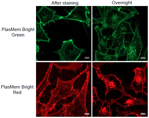
- ・PlasMem Bright Green
(Dye concentration: 200-fold diluted)
(Ex: 488 nm, Em: 500–560 nm)
・PlasMem Bright Red
(Dye concentration: 100-fold diluted)
(Ex: 561 nm, Em: 560–700nm)
Figure 4. Images of plasma membrane fluorescence from PlasMem Bright dyes in HeLa cells cultured for 1 day.
Detection of fluorescent mitochondria in nerve axons.
- SH-SY5Y cells (2.4x 10⁵ cells/ml) in DMEM/F11 (containing 10% fetal bovine serum, 1% penicillin-streptomycin) were seeded on a μ-slide 8-well plate (Ibidi) and cultured overnight in an incubator set at 37 °C and equilibrated with 95% air and 5% CO2.
- After the supernatant was removed, DMEM/F11 medium (10 μmol/L retinoic acid, 1% FBS) was added, and the cells were cultured for 7 days in an incubator set at 37 °C and equilibrated with 95% air and 5% CO2. The medium was replaced with fresh medium every 2 days.
- The supernatant was replaced with a working solution (Hoechst: 2 μg/ml, PlasMem Bright Green: 200-fold or PlasMem Bright Red: 100-fold, MitoBright LT: 0.1 μM, 200 μl), and the cells were cultured for 15 minutes in an incubator set at 37 °C and equilibrated with 95% air and 5% CO2.
- The cells were washed twice with 200 μl of HBSS.
- DMEM/F11 medium was then added, and the cells were observed under a fluorescence microscope.
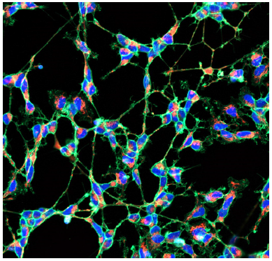
- (Blue) Hoechst 33342: 405 nm (Ex), 400-450 nm (Em)
(Green) PlasMem Bright Green: 488 nm (Ex), 500–560 nm (Em)
(Red) MitoBright LT Red: 561 nm (Ex), 560–620 nm (Em)
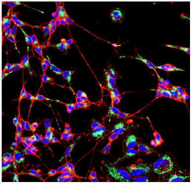
- (Blue) Hoechst 33342: 405 nm (Ex), 400-450 nm (Em)
(Green) MitoBright LT Green: 488 nm (Ex), 500–560 nm (Em)
(Red) PlasMem Bright Red: 561 nm (Ex), 560–700 nm (Em)
Figure 5. Multi-color images of plasma membrane and mitochondria in the differentiated SHSY-5Y cells
stained with PlasMem Bright dyes and MitoBright LT dyes.
References
- Shi, L., et al., Angewandte Chem. Int. Ed., 2020, 59, 9962-9966.
- Collot, M., et al., Bioconjugate Chem., 2020, 31(3), 875-883.
- Shimomura, T., et al. bioRxiv, 2020, doi: https://doi.org/10.1101/2020.02.02.931295.
P504_P505: PlasMem Bright Green/PlasMem Bright Red
Revised Jun., 26, 2023