 Hidden sections will not be printed.
General Information
Mitochondrial respiration is the process by which cells synthesize adenosine triphosphate (ATP), the cellular source of energy. As part of the respiration process, the electron transport chain generates the proton-motive force. The electron transport chain consists of five enzyme complexes (complexes I–V), with complex I (NADH: ubiquinone oxidoreductase) acting as the entry point. Through electron transfer from NADH to ubiquinone, complex I transports protons from the mitochondrial matrix to the intermembrane space. Complex I is implicated in neurodegenerative diseases such as Parkinson's disease, mitochondrial disorders, cancer, and aging. It is considered one of the key enzyme complexes for analyzing mitochondrial function, elucidating disease mechanisms, and developing therapeutic agents.
MitoComplex-I Activity Assay Kit enables the determination of complex I activity in isolated mitochondria by measuring NADH oxidation. This kit is designed for use with 96-well microplates, making it suitable for the simultaneous processing of multiple samples, such as compound screening or the evaluation of drugs targeting complex I. When measuring Complex I activity in mitochondria fractionated using the IntactMito Fractionation Kit for Tissue (Code: MT17, Dojindo), please use this kit.
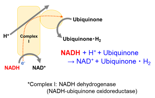 |
| Figure 1. Principle of the MitoComplex-I Activity Assay Kit |
Kit Contents
| Assay Buffer | 25 ml × 1 |
| Assay Reagent | × 1 |
| Coenzyme | × 1 |
| Ubiquinone | × 1 |
Storage Conditions
Store at 0–5 ℃
Required Equipment and Materials
- Ethanol
- Plate reader (340 nm filter)
- 96-well plate
- 10 μl and 100 - 200 μl multi-channel pipettes
Precautions
- Equilibrate reagents to room temperature prior to use.
- Briefly centrifuge the tubes before opening to ensure the content is at the bottom.
- Analysis of samples in triplicate is recommended for accuracy.
Preparation of Solutions
Preparation of Assay stock solution
Add 1200 µl of double-distilled H2O (ddH2O) to the Assay Reagent tube and dissolve by gentle pipetting.
- Assay stock solution is stable for 1 month when stored at -20 ℃.
Preparation of Coenzyme stock solution
Add 110 µl of ddH2O to the Coenzyme tube and dissolve by gentle pipetting.
- Coenzyme stock solution is stable for 1 month when stored at -20 ℃.
Preparation of Ubiquinone stock solution
Add 100 µl of ethanol to the Ubiquinoe tube and dissolve by gentle pipetting.
- Ubiquinone stock solution is stable for 1 month when stored at -20 ℃.
Preparation of the Working solution
Add the required amounts of Assay Reagent Stock Solution, Coenzyme Stock Solution, Ubiquinone Stock Solution, and Assay Buffer to a tube, and mix well by gentle pipetting.
- Prepare sufficient volume for three wells each containing a sample and a blank, as needed.
- Use the prepared Working solution on the same day.
Solution preparation example
| 6 well | 96 well | |
| Assay reagent stock solution | 80 μl | 1200 μl |
| Coenzyme stock solution | 6.4 μl | 96 μl |
| Ubiquinoe stock solution | 6 μl | 90 μl |
| Assay Buffer | 307.6 μl | 4614 μl |
General Protocol
- Add 25 μl per well of the protein concentration-adjusted mitochondrial suspension to a 96-well microplate.
- The recommended mitochondria are those that have been frozen for at least one night. For more information, please refer to the FAQ section on our product website.
- The recommended protein concentration of the mitochondrial suspension is 5–20 μg per well.
- Dilute the mitochondrial suspension with Assay Buffer and mix by gentle pipetting.
- Add 25 μl of each inhibitor solution or Assay Buffer per well.
- Prepare inhibitor solutions using Assay Buffer.
- Add only Assay Buffer to the control wells.
- Add 50 μl per well of the Working solution to each well.
- Add the solution gently to avoid bubbles.
- Immediately measure absorbance at 340 nm using a microplate reader (25 ℃, 1-minute intervals for 10-30 minutes).
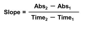
- Select the linear range of the reaction from the start of the measurement and calculate the rate of change in absorbance (slope) within this interval as the enzyme activity.
- To accurately measure Complex I activity, it is recommended to compare results with those obtained in the presence of Rotenone, a Complex I inhibitor.
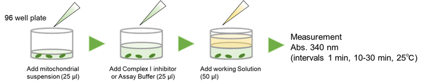 |
| Figure 2. Procedure for the measurement of Complex I activity using MitoComplex-I Activity Assay Kit |
Experimental Example
Measurement of Complex I Activity in Mitochondria that were fractionated using the IntactMito Fractionation Kit for Tissue (Code: MT17)
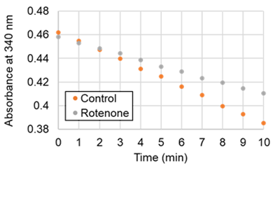
- Mitochondria were fractionated from mouse brain tissue using the IntactMito Fractionation Kit for Tissue (Code: MT17), and the mitochondrial suspension(protein concentration: 0.8 mg/ml, 25 μl/well)was added to a 96-well microplate.
- The mitochondrial suspension was diluted gently with Assay Buffer by pipetting to achieve a final protein concentration of 0.8 mg/ml.
- Assay Buffer (25 μl) was added to the well of Control, and Rotenone (10 μmol/l, 25 μl) prepared with Assay Buffer was added to the well of Rotenone.
- Working solution (50 μl) was added to each well of the Control and Rotenone.
- The solution was added gently to avoid bubbles.
- Absorbance at 340 nm was measured immediately using a microplate reader (25 ℃, 1-minute intervals for 10 minutes).
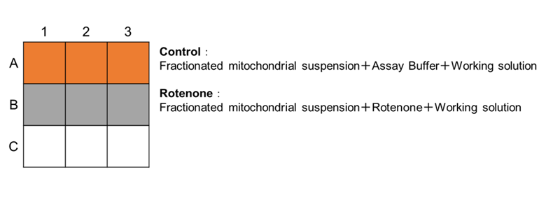 |
| Figure 3. Plate layout |
 |
 |
|
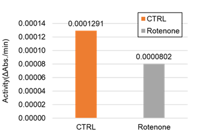
| Figure 4. Absorbance change from 0 to 10 min | Figure 5. Complex I activity |
|
The rate of absorbance changes at 340 nm from 0 to 10 min were calculated, and the complex I activity was determined to be 0.00244 (ΔAbs./min/mg protein) using the following equation.
Complex I Activity = {Control (ΔAbs./min) – Rotenone (ΔAbs./min)}/protein (mg)
Frequently Asked Questions / Reference


MT18: MitoComplex-I Activity Assay Kit
Revised Nov., 25, 2025