 Hidden sections will not be printed.
General Information
Mitochondria are the principal cellular organelle for oxidative phosphorylation and the production of ATP and mitochondrial dysfunction is relevant to cancer, cell senescence and neurodegenerative diseases such as Alzheimer’s and Parkinson’s.
Methods for monitoring mitochondrial morphology, dynamics, and number are usually based on small fluorescent molecules or plasmid transfection techniques. The use of plasmids requires the target protein to be stably expressed, while small fluorescent molecules are widely used because they can simply be added to cells. Among commercially available small fluorescent molecules, those containing the chloromethyl moiety are commonly used. However, these dyes have some limitations, including short-term retention in cells, decreased fluorescence intensity in serum, and high background.
Dojindo’s MitoBright LT dyes overcome these limitations. MitoBright LT dyes are designed to exhibit mitochondrial retention for long-term visualization. In addition, the MitoBright LT dyes show stronger fluorescence signals compared with other commercially available dyes that contain the chloromethyl moiety. The MitoBright LT dyes offer three different color options (Green, Red and Deep Red), and are provided as a ready-to-use DMSO solution. A working solution can easily be prepared in a single dilution step with growth medium or HBSS.
Contents
Staining number possible by Unit Size (35 mm dish)
| Unit size : 400 µl | |
| MT10 : MitoBright LT Green | 200 |
| MT11 : MitoBright LT Red | |
| MT12 : MitoBright LT Deep Red |
Storage Condition
| MT10 : MitoBright LT Green | Store at -20℃, protected from light and moisture. |
| MT11 : MitoBright LT Red | Store at -20℃, protected from light and moisture. |
| MT12 : MitoBright LT Deep Red | Store at -20℃, protected from light and moisture. |
Required Equipment and Materials
- Growth medium or HBSS
- Micropipettes
Preparation of Solutions
Preparation of MitoBright LT working solution
Dilute the 0.1 mmol/L MitoBright LT solution with growth medium to prepare a 0.1 μmol/L MitoBright LT working solution.
- Please use the MitoBright LT working solution within the day.
General protocol
- Seed cells in a dish and culture them overnight in a 37℃ incubator equilibrated with 95% air and 5% CO2.
- Discard the supernatant and wash the cells once with growth medium.
- Add the 0.1 μmol/l MitoBright LT working solution to the cells and incubate them in a 37℃ incubator equilibrated with 95% air and 5% CO2 for 15 minutes.
- Discard the supernatant and wash the cells twice with growth medium.
- Add growth medium or HBSS to the cells, then observe the cells under a fluorescence microscope.
Usage Examples
Detection of fluorescent mitochondria in HeLa cells over time
- HeLa cells in MEM (containing 10% fetal bovine serum, 1% penicillin-streptomycin) were seeded on a μ-slide 8 well plate (ibidi) and cultured overnight in a 37℃ incubator equilibrated with 95% air and 5% CO2.
- After the supernatant was removed, MitoBright LT working solution (0.1 μmol/l, 200 μl) was added and the cells were cultured in a 37℃ incubator equilibrated with 95% air and 5% CO2 for 30 minutes.
- The cells were then washed twice with 200 μl MEM.
- MEM (without phenol red) was then added, and the cells were observed over time under a fluorescence microscope.
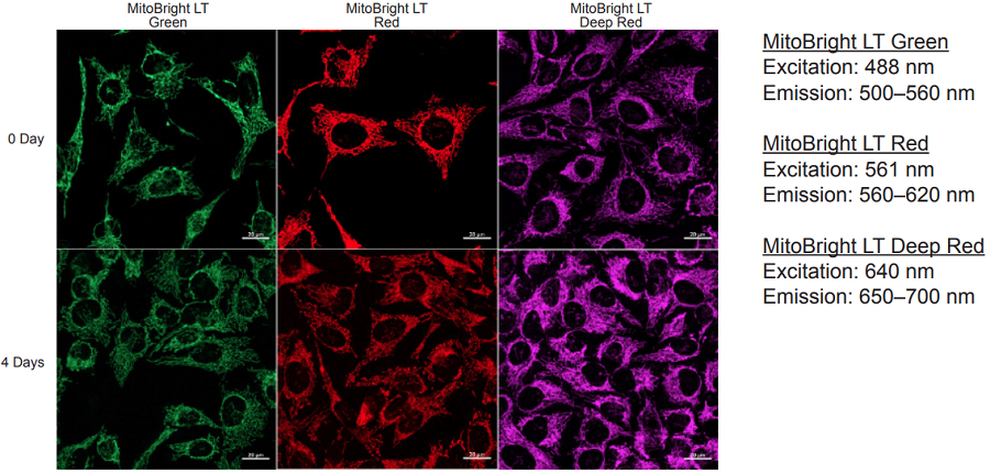
Figure 1 Images of mitochondrial fluorescence using MitoBright LT dyes in cells cultured for 4 days.
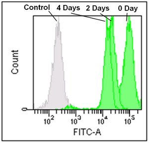
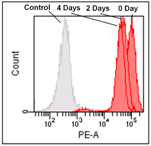
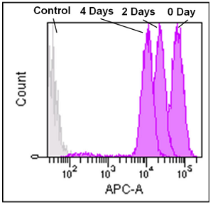
Flow cytometry analysis of mitochondria in Jurkat cells over time
- Jurkat cells in RPMI (containing 10% fetal bovine serum, 1% penicillin-streptomycin) were seeded in a dish and were cultured overnight in a 37℃ incubator equilibrated with 95% air and 5% CO2.
- After the supernatant was removed, MitoBright LT working solution (0.1 μmol/l, 5 ml) was added, and the cells were cultured in a 37℃ incubator equilibrated with 95% air and 5% CO2 for 30 minutes.
- The cells were then washed twice with 5 ml RPMI.
- RPMI was then added and the cells were analyzed over time by flow cytometry.
MitoBright LT Green
Ex: 488 nm
Em: 515 nm–545 nm
MitoBright LT Red
Ex: 488 nm
Em: 564–604 nm
MitoBright LT Deep Red
Ex: 633 nm
Em: 650–670 nm
Figure 2 Flow cytometry analysis of mitochondria over time using MitoBright LT dyes.
Excitation and emission spectra of MitoBright LT dyes
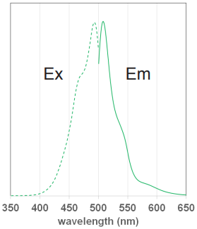
Figure 3 Excitation and emission
spectra of MitoBright LT Green
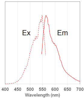
Figure 3 Excitation and emission
spectra of MitoBright LT Green
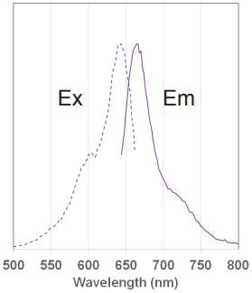
Figure 5 Excitation and emission
spectra of MitoBright LT Deep Red
Fluorescence imaging of mitochondria using MitoBright LT dyes
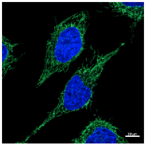
Concentration of dye: 0.1 μmol/L
Cell line: HeLa cells
Excitation: 488 nm
Emission: 500–560 nm
Nuclear stain: Hoechst 33342
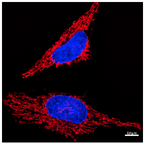
Concentration of dye: 0.1 μmol/L
Cell line: HeLa cells
Excitation: 561 nm
Emission: 560–620 nm
Nuclear stain: Hoechst 33342
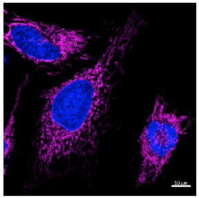
Concentration of dye: 0.1 μmol/L
Cell line: HeLa cells
Excitation: 640 nm
Emission: 650–700 nm
Nuclear stain: Hoechst 33342
MT10_MT11_MT12: MitoBright LT Green / Red / Deep Red
Revised Jun., 21, 2023