 Hidden sections will not be printed.
General Information
Ab-10 Rapid HiLyte FluorTM 555 Labeling Kit enables rapid (in less than 30 min) and easy labeling of HiLyte FluorTM 555 to 10 µg antibody. Reactive HiLyte FluorTM 555 (a component of the kit) has succinimidyl ester group, that can easily make a covalent bond with an amino group of the target antibody without any activation process. This kit contains all the necessary reagents to prepare a HiLyte FluorTM 555-labeled antibody.
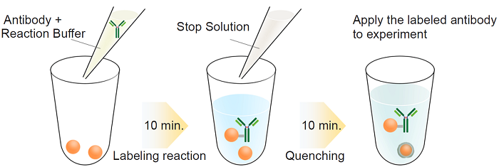
Fig.1 Labeling procedure
|
Caution After a Reactive HiLyte FluorTM 555 is taken out from the seal bag, keep the unused Reactive HiLyte FluorTM 555 in the bag, seal tightly and store at -20°C. Store the other components at 0-5°C. |
Kit Contents
| Reactive HiLyte FluorTM 555 | x 3 |
| Reaction Buffer | 100 µl x 1 |
| Stop Solution | 100 µl x 1 |
Storage Condition
Store at 0-5°C
This kit is stable for 1 year at 0-5°C before opening.
Required Equipment and Materials
- 20 µl adjustable pippet
- Microtube (for sample preparation)
- Incubator (37°C)
- PBS (Phosphate buffered saline)
Precaution
- Use 0.5-1 mg/ml of antibody solution for labeling. If the antibody concentration is more than 1 mg/ml, please dilute the antibody solution with PBS.
- If the sample solution contains small insoluble materials, centrifuge the solution, and use the supernatant for the labeling.
- The microtubes in this kit contain solutions. Since there is a possibility that the droplets might attach to the inside walls or caps, please spin the tube to drop them down prior to open.
- In case an antibody solution includes a high concentration of constituents, such as BSA or glycerol, it may interfere with a labeling and cause a non-specific signal. We recommend removing the constituents prior to labeling procedure. Usable constituents (○) and non-usable constituents (×) are shown in Table 1, and compatible concentrations of constituents are shown in Table 2.
| Additives | |
| Buffering agents (PBS,HEPES) | ◯ |
| Sodium chloride | ◯ |
| Chelating agents (EDTA) | ◯ |
| Sodium azide | ◯ |
| Primary amines and thiols | X |
| Glucose | Glycerol | BSA | Gelatin | Tris | |
| Anti-Mitochondria antibody | < 10% | < 10% | < 2% | < 0.1% | < 50mmol/L |
| Anti-Actin antibody | < 5% | < 10% | X | < 0.1% | < 25mmol/L |
| Anti-HNF4α antibody | < 2% | < 10% | < 0.05% | < 0.02% | < 50mmol/L |
- Interference and non-specific signal may be dependent on types of antigen, host species of antibody or constituents.
Protocol
- Add 0.5-1 mg/ml of the antibody solution to a microtube to be an amount of antibody of 10 µg.
- Add Reaction Buffer to the antibody solution (step 1) and mix by pipetting.
- The volume of Reaction Buffer: one-tenth of the antibody solution (Table 3).
- Add the solution (step 2) to Reactive HiLyte FluorTM 555 and mix by pipetting.
- Incubate at 37°C for 10 minutes.
- Add Stop Solution to the solution (step 4) and mix by pipetting.
- The volume of Stop Solution: one-tenth of the antibody solution (Table 3).
- Incubate at room temperature for 10 minutes.
- Apply the sample (step 6) for desired experiments or store at 0-5 °C.
- The labeled antibody is stable at 4°C for 2 weeks.
| The concentration of antibody (mg/ml) | 0.5 | 0.6 | 0.7 | 0.8 | 0.9 | 1.0 |
| The volume of Reaction Buffer (μl) | 2.00 | 1.67 | 1.43 | 1.25 | 1.11 | 1.00 |
| The volume of Stop Solution (μl) | 2.00 | 1.67 | 1.43 | 1.25 | 1.11 | 1.00 |
Supplimental Information
Mitochondria immunostaining
- HeLa cells were seeded on a µ-slide 8 well (ibidi) and cultured overnight at 37 ℃ in a 5% CO2 incubator.
- The cells were washed with PBS three times, and 4% paraformaldehyde in PBS was added to the µ-slide.
- The cells were then incubated at room temperature for 15 minutes.
- The cells were washed with PBS three times, and 1% Triton-X in PBS was added to the µ-slide.
- The µ-slide was incubated at room temperature for 30 minutes.
- Once the cells were washed with PBS three times, a blocking solution prepared with PBS was added to the µ-slide.
- The cells were then incubated at room temperature for 1 hour.
- HiLyte FluorTM 555 conjugated anti-mitochondria antibody was diluted 200 times with the blocking solution.
- Anti-mitochondria antibody was purchased from Abcam (Product Code: ab3298).
- The supernatant was discarded and the solution (step 8) was added to the µ-slide.
- The µ-slide was incubated at 0-5℃ overnight.
- After the cells were washed with PBS-T three times, PBS-T was added to the µ-slide.
- The cells were observed under a fluorescence microscope.
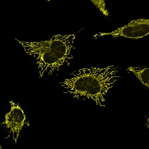 Fig. 2 Microscope image of mitochondria in HeLa cells |
Frequently Asked Questions / Reference


LK35: Ab-10 Rapid HiLyte Fluor™ 555 Labeling Kit
Revised Dec., 05, 2023