 Hidden sections will not be printed.
Kit Contents
| Reactive R-Phycoerythrin | x 3 |
| Reaction Buffer | 100 µl x 1 |
| Stop Splution | 100 µl x 1 |
Storage Condition
Store at 0-5°C
This kit is stable for 1 year at 0-5°C before opening.
Required Equipment and Materials
- 20 µl adjustable pippet
- Microtube (for sample preparation)
- Incubator (37°C)
- PBS (Phosphate buffered saline)
General Information
Ab-10 Rapid R-Phycoerythrin labeling Kit is rapid (in less than 30 min) and easy preparation kit of R-Phycoerythrin-labeled antibody (Ab) for 10 µg antibody. Reactive R-Phycoerythrin (a component of the kit) has succinimidyl ester groups, that can easily make a covalent bond with an amino group of the target antibody without any activation process. This kit contains all the necessary reagents to prepare a R-Phycoerythrin-labeled antibody.
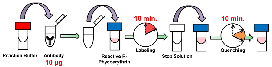
Fig 1. Labeling procedure
|
Caution After a Reactive R-Phycoerythrin is taken out from the seal bag, keep the unused Reactive R-Phycoerythrin(s) in the bag, seal tightly and store at -20°C. Store the other components at 0-5°C. |
Precaution
- Use 0.5-1 mg/ml of antibody solution fo labeling. If the antibody concentration is more than 1 mg/ml, please dilute the antibody solution with PBS.
- If the sample solution contains small insoluble materials, centrifuge the solution, and use the supernatant for the labeling.
- The microtubes in this kit contain solutions. Since there is a possibility that the droplets might attach to the inside walls or caps, please spin down to drop them down prior to open.
- Some additives in an antibody solution may interfere with the labeling if the concentration is too high. The maximum compatible concentrations of such additives are indicated in Table 1.
|
|
- Containing BSA may result in non-specific signal depending on the antibodies used. Removing BSA prior to the R-Phycoerythrin labeling is recommended in case high non-specific signalis observed.
Protocol
- Add 0.5-1 mg/ml of the antibody solution to a microtube to be an amount of antibody of 10 µg.
- Add Reaction Buffer to the antibody solution (step 1) and mix by pipetting.
- The volume of Reaction Buffer: one-tenth of the antibody solution (Table 2).
- Add the solution (step 2) to Reactive R-Phycoerythrin and mix by pipetting.
- Incubate at 37°C for 10 minutes.
- Add Stop Solution to the solution (step 4) and mix by pipetting.
- The volume of Stop Solution: one-tenth of the antibody solution (Table 2).
- Incubate at room temperature for 10 minutes.
- Apply the sample (step 6) for desired experiments or store at 0-5°C.
- The labeled antibody is stable at 4°C for 2 weeks.
| The concentration of antibody (mg/ml) |
0.5 | 0.6 | 0.7 | 0.8 | 0.9 | 1.0 |
| The volume of Reaction Buffer (µl) |
2.00 | 1.67 | 1.43 | 1.25 | 1.11 | 1.00 |
| The volume of Stop Solution (µl) |
2.00 | 1.67 | 1.43 | 1.25 | 1.11 | 1.00 |
Supplimental Information
Fluorescent staining HL 60 cells
- HL60 cells were added to the number of 5 x 10⁵ cells/tube in a microtube.
- The supernatant was removed by centrifugation at 1,000 x g for 2 minutes.
- Suspension buffer [50 µl, 1% FBS (fetal bovine serum), Hank's HEPES balanced buffer] was added to the tube.
- R-Phycoerythrin conjugated antibody (1 µg) was added to the tube and mixed by pipetting.
- Anti-CD13 antibody was purchased from Becton Dickinson(Product Code: 555393). Mouse IgG (Isotype) was purchased from Jackson Immuno Research Laboratories (Product Code: 015-000-003).
- The tube was incubated on ice for 30 minutes.
- The supernatant was removed by centrifugetion at 1,000 x g for 2 minutes.
- Suspension buffer (1 ml) was added to the tube and the cells were suspended by pipetting.
- Stained cells were analysed by flow cytometry.
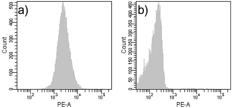
Fig.2 Staining HL60 cells with R-Phycoerythrin labeledantibody |
Frequently Asked Questions / Reference


LK34: Ab-10 Rapid R-Phycoerythrin Labeling Kit
Revised Feb., 25, 2026