 Hidden sections will not be printed.
General Information
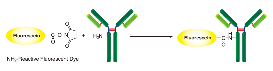
Fluorescein Labeling Kit - NH2 is primarily used for the preparation of fluorescein-labeled antibody for immunostaining and cellular proteins for tracing. NH2-Reactive Fluorescein, a component of this kit, has a succinimidyl ester group, and can easily make a covalent bond with an amino group of the target protein or other macromolecules without any activation process. Filtration Tube included in this kit is used for sample protein in removing small molecules such as Tris buffer and amine compounds that interfere with the assay or labeling reaction. The labeling process is very simple. Add the NH2-Reactive Fluorescein to protein solution on a filter membrane, and incubate at 37oC for 10 min. Excess Fluorescein molecules can be removed by a filtration tube. The excitation and emission wavelengths of the fluorescein-labeled proteins are 500 nm and 525 nm, respectively. This kit contains the necessary reagents for labeling, including the storage buffer for conjugates.
Kit Contents
| NH2-Reactive Fluorescein | 3 tubes |
| WS Buffer | 4 ml x 1 |
| Reaction Buffer | 500 μl x 1 |
| Filtration Tube | 3 tubes |
Capacity
Three samples labeling
- Sample requirement: Molecular weight > 50,000; amount: 50-200 μg
Storage Condition
Store at 0-5oC. This kit is stable for 1 year at 0-5oC before opening.
|
Caution After a NH2-Reactive Fluorescein is taken out from the seal bag, keep the unused NH2-Reactive Fluorescein(s) in the bag, seal tightly and store at -20oC. |
Required Equipment
- 10 μl and 200 μl adjustable pipettes
- Incubator (37oC)
- Microtubes
- Microcentrifuge
- DMSO
Precaution
- If the target protein solution contains other proteins with molecular weight larger than 10,000 such as serum albumin or gelatin, purify the protein solution, and use the purified target proteins for fluorescein labeling, because it might interfere the labeling reaction.
- If the protein solution contains small insoluble materials, centrifuge the solution, and use the supernatant for the labeling.
- The droplets which induced from the dry inhibitor of membrane, are occasionally found inside Filtration Tube while storing the kit at 0-5oC or after returning to room temperature. This phenomenon does not affect the performance.
General Protocol
-
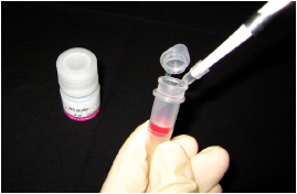
Step 1.
Add 100 μl WS Buffer and the sample solution containing 50-200 μg proteina) to a Filtration Tube.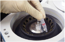
Step 2.
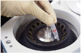
Mix the solution with pipetting several times, and centrifuge at 8,000 x g for 10 min.b)
-
Step 3.
Add 10 μl DMSO to NH2-Reactive Fluorescein, and dissolve with pipetting.c)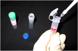
Step 4.
Add 100 μl Reaction Buffer to the Filtration Tube, and then add 8 μl d) NH2-Reactive Fluorescein solution to the Filtration Tube and pipette to mix
-
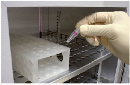
Step 5.
Incubate the tube at 37°C for 10 min.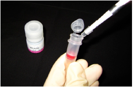
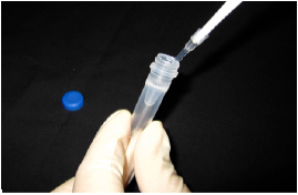
Step 6.
Add 100 μl WS Buffer to the Filtration Tube, and centrifuge at 8,000 x g for 10 min.b) Discard the filtrate.
-
Step 7.
Add 200 μl WS Buffer to the Filtration Tube, and centrifuge at 8,000 x g for 10 min.b) Repeat this step one more time.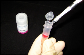
Step 8.
Add 200 μl WS Buffer, and pipette about 10 times to recover the conjugate.e) Transfer the solution to a microtube (not included in this kit), and store at 0-5°C.
a) The volume of protein solution should be less than 100 μl. If the protein concentration is lower than 0.5 mg/ml, repeat Steps 1 and 2 until the total protein accumulation becomes 50 - 200 μg.
b) If the solution still remains on the membrane after the centrifugation, spin for another 5 min.
c) NH2-Reactive Fluorescein is on the bottom of the tube. Add 10 μl DMSO to the bottom of the tube, and pipette several times to dissolve.
NH2-Reactive Fluorescein can be hydrolyzed by moisture in DMSO. Proceed to Step 4 immediately after the preparation of the NH2-Reactive Fluorescein solution.
d) If the amount of protein is 200 μg, add entire NH2-Reactive Fluorescein solution.
e) We recommend using WS Buffer to recover the conjugate. You can choose any kinds of buffers appropriate for your experiment.
Determination of Fluorescein / protein Ratio
Dilute the fluorescein-labeled protein solution with WS Buffer or other neutral buffer to a proper volume then measure the absorbance of the protein solution at 280 nm and 500 nm, if you need to know the fluorescein/protein ratio. Calculate the ratio using the following equation: When target protein is IgG, use 216,000 as the ε of IgG. Molar absorption coefficient of fluorescein in WS Buffer is 60,000.
-
Ratio (fluorescein molecules per protein molecule) = A500 / 60,000 (A280 - A500 x 0.22) / (ε of protein)
- A500: absorbance at 500 nm
A280: absorbance at 280 nm
ε: molar absorption coefficient of protein at 280 nm
Fluorescein Labeling Reaction
Q & A
Can I use this kit to label antibodies which is commercially available?
Yes. However, if antibody solution contains other proteins such as serum albumin or gelatin, labeling reaction might be interefered by thatprotein. Purification of the antibody solution with affinity chromatography is necessary prior to use this kit.
Contact us for the purification procedure, if you need.
How many fluorescein molecules are introduced to protein?
The number of conjugated fluorescein depends on the protein. In the case of rabbit IgG, 3 to 7 fluorescein molecules conjugate to each protein molecule.
How long is the fluorescein-labeled protein stable?
Stability of conjugate depends on the protein itself. In the case of labeling for rabbit IgG, the labeled IgG is stable at 4oC for 2 months. For longer storage, add equal volume of glycerol to the sample solution and store at -20oC.
What is a minimum amount of protein that can be labeled using this kit?
We recommend using 50 μg as a minimum amount. Though 10 μg protein can be labeled using this kit, the background might be increased.
Can I use this kit to label oligonucleotides or oligopeptides?
No. Oligonucleotides and oligopeptides may be too small to retain on the membrane filter of Filtration Tube.
Is there any notice for treatment of living cells with the fluorescein-labeled protein?
We recommend using PBS including 2-10% FBS for preparation of cell suspension to maintain the best cell conditions.
Does recovery buffer (WS Buffer) have harmful effect to living cells?
No. WS Buffer contains stabilizing agent (surfactant) that is controlled of its concentration without cytotoxicity. If you are concerned about the additive in WS Buffer, you can use your own buffer currently used instead of WS Buffer.
Frequently Asked Questions / Reference


LK01: Fluorescein Labeling Kit - NH2
Revised Jan., 05, 2024