 Hidden sections will not be printed.
General Information
Iron is one of the most important metallic elements in living organisms. In recent years, an iron-dependent mode of cell death, ferroptosis, has emerged, and research on intracellular iron ions has attracted increased attention.
The Iron Assay Kit-Colorimetric is a kit for measuring the amount of iron ions (Fe²⁺ and Fe³⁺) in tissue. It enables data acquisition in approximately 2 h from sample pretreatment to the measurement of iron ion concentration.
The concentration of Fe²⁺ in a tissue sample can be determined by adding Ferene S, which is a chromogenic dye for Fe²⁺, to the tissue lysate and measuring its absorbance. By using the reducing agent included in the kit to reduce Fe³⁺ in the sample to Fe²⁺, the concentrations of total iron and Fe³⁺ in the sample can also be determined.
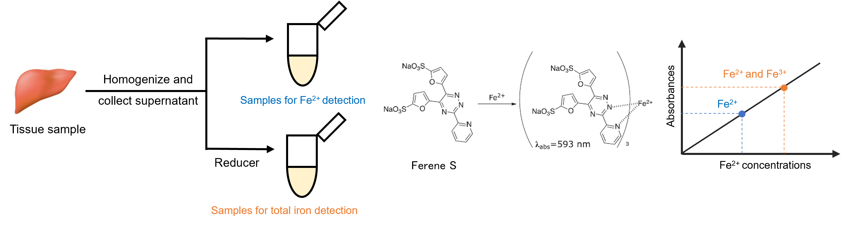
Figure 1. Principle of the Iron Assay Kit-Colorimetric
Kit Contents
| 50 tests | |
| Probe Solution | 7 ml× 1 |
| Assay Buffer | 20 ml× 1 |
| Standard (Yellow cap) | 150 μl (1 mmol/l)× 1 |
| Reducer (Blue cap) | × 1 |

Approximate number of detectable samples for reference (n=3)
Storage Conditions
Store at 0–5 °C and protect from light.
Required Equipment and Materials
- Homogenizer or mortar and pestle
- 20–200 µl multichannel pipette
- 100–1000 µl, 20–200 µl micropipette
- 1.5 ml microtube
- PBS (-)
- Ice bath
- Centrifuge
- Micro plate reader (593 nm)
- 96-well microplate
- Ultrasonic bath
Precautions
- Bring all reagents in the kit to room temperature before use.
- Due to vibration during transport, contents may adhere to the inner walls of the tubes or to the undersides of the tube caps. Ensure they settle at the bottom of the tubes before opening.
- To obtain accurate results, use tissue as fresh as possible and keep the sample on ice during preparation.
- For accurate measurement, use multiple wells (n = 3 or more) per sample.
- For samples with a high iron content, dilute with assay buffer for measurement to ensure the final concentration falls within the range of the standard curve.
Preparation of Solutions
Preparation of Reducer solution
1. Add 300 µl of Assay Buffer to Reducer and sonicate for approximately 1 minute to completely dissolve the contents.
- Reducer solution can be stored at 0-5°C for up to 2 months.
Preparation of tissue sample
1. Weigh a fresh tissue sample (e.g., 100 mg) into a 1.5 ml microtube.
2. Add cold PBS (-) and wash the sample by pipetting. Centrifuge at 16,000×g for 4 minutes and remove the supernatant.
3. Add 1.3 ml of Assay Buffer and homogenize the sample in an ice bath.
4. Sonicate the microtube containing the sample in an ultrasonic bath for 5 minutes.
- To prevent the sample temperature from rising, immerse the microtube in ice bath for 20 seconds every minute.
5. Centrifuge at 16,000×g for 10 minutes and transfer the supernatant to a new 1.5 ml microtube.
6. Transfer 400 µL of the collected supernatant to each of three new 1.5 ml microtubes and prepare the ferrous iron, total iron, and sample blank solutions.
Preparation of Standard solution
1. Add 100 μL of Standard (1 mmol/l) to a 1.5 ml microtube containing 900 μl of assay buffer to prepare a 100 μmol/l standard solution.
2. As shown in Figure 2, add 400 µl of assay buffer to each 1.5 ml microtube. Add 400 µl of 100 µlmol/l standard solution, mix, and then transfer 400 µl to the next tube. Repeat this procedure to prepare standard solutions of 50, 25, 12.5, 6.25, 3.125, 1.563, and 0.781 µmol/l. Also, prepare a solution of 0 µmol/l (assay buffer only).
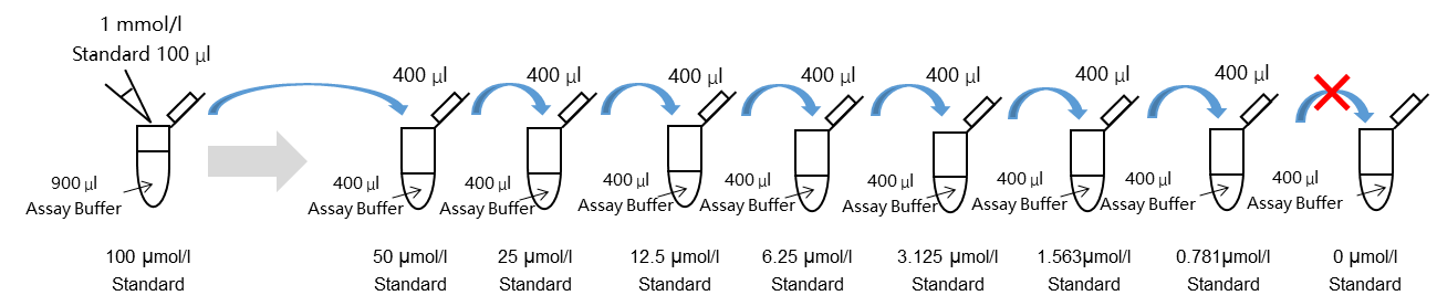
Figure 2. Preparation of Standard solution
General Protocol
1. Add reducer solution to each standard solution at a 5% volume ratio. For example, add 20 µl of reducer solution to each 400 µl of standard solution (50–1.563, 0 μmol/l), and add 40 μl of reducer solution to 800 µl of standard solution (0.781 μmol/l).
2. Add Assay Buffer at 5% volume to each Fe²⁺ sample solution and sample blank solution. For example, add 20 μl of assay buffer to 400 μl of Fe2+ sample solution or sample blank solution.
3. Add reducer solution at 5% volume to the total iron sample solution. For example, add 20 μl of reducer solution to 400 μl of total iron sample solution.
4. Incubate all solutions prepared in Steps 2 and 3 in an incubator at 37°C for 15 minutes.
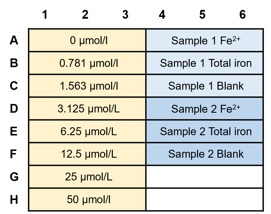
5. Add 105 μl of the solutions prepared in Step 4 to the wells of a 96-well plate (refer to Tables 1 and 2).
6. Add 100 μl of probe solution to each well containing standard solution, Fe2+ sample solution, or total iron sample solution. Add 100 μl of assay buffer to wells containing the sample blank solution.
7. Incubate at 37 °C for 1 hour.
8. Measure the absorbance at 593 nm using a microplate reader.
Table1. Amount of each solution for standard, sample, blank well

Table 2. Example of standard, sample, and blank arrangement on a 96-well plate (n=3)
Experimental Example
Measurement of Fe2+ and Fe3+ concentrations in mouse liver tissue
1. Liver tissue (100 mg) was weighed into a 1.5 ml microtube.
2. Cold PBS (1 ml) was added, and the sample was washed 20 times by pipetting. Then, it was vortexed for 10 seconds and centrifuged at 16,000×g for 4 minutes before removing the supernatant.
3. The sample was transferred to a mortar, frozen with liquid nitrogen, and thoroughly ground with a pestle until fully powdered. Assay buffer (1.3 ml) was added, and the suspension was transferred to a microtube.
4. The microtube containing the sample was sonicated in an ultrasonic water bath for 5 minutes; to prevent the temperature of the sample from rising, it was placed in an ice bath for 20 seconds every minute.
5. The tube was centrifuged at 16,000×g for 10 minutes, and the supernatant was transferred to a new 1.5 ml microtube.
6. The collected supernatant was transferred to three new 1.5 ml microtubes (400 µl in each tube) to prepare a ferrous iron sample solution, a total iron sample solution, and a sample blank solution.
7. Standard solutions were prepared as described in the section “Preparation of Standard solution."
8. Reducer solution (20 µl) was added to both the standard solution and the total iron sample solution, and Assay buffer (20 µl) was added to the Fe²⁺ sample solution and the sample blank.
9. The solutions prepared in Step 8 were incubated at 37°C for 15 minutes.
10. The solutions (105 µl) prepared in Step 9 and probe solution (100 µl) were added to the wells of a 96-well plate (refer to Tables 1 and 2), and incubated at 37°C for 1 hour.
11. The absorbance at 593 nm was measured using a microplate reader, and the concentrations of Fe2+ and Fe3+ in the samples were calculated from the calibration curve.
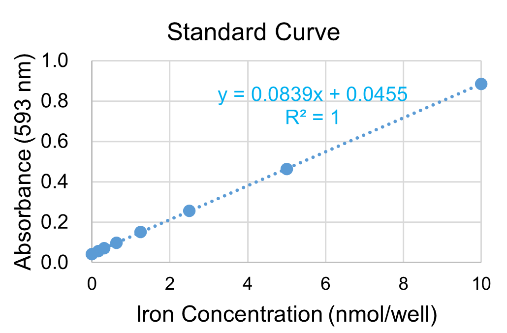
Figure 3. Standard curve
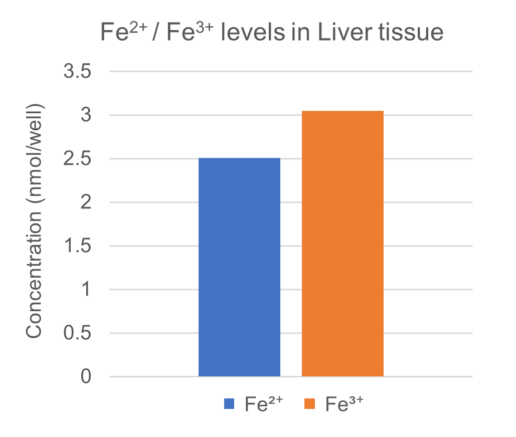
Figure 4. Measurement of the concentration of Fe2+ and Fe3+ in liver tissue
I291: Iron Assay Kit -Colorimetric-
Revised Sep., 25, 2025