 Hidden sections will not be printed.
General Information
H2AX is phosphorylated rapidly and widely upon DNA double-strand breaks. Among H2AX histone variants, H2AX with phosphorylated Ser-139 is called γH2AX. As a DNA damage marker, γH2AX is monitored to investigate genotoxicity and carcinogenicity of chemicals. Recent studies suggest that γH2AX is a marker for cellular senescence. The DNA Damage Detection Kit includes all necessary components, an anti-γH2AX antibody, secondary antibody (fluorophore labeled), and blocking solution for staining which allows ready-to-use immunodetection of γH2AX.

Figure 1. Detection of γH2AX
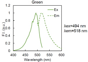
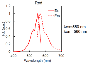
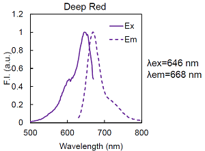
Figure 2. Excitation and emission spectra of fluorophores
Kit Contents
|
Anti γH2AX antibody (Host ; Mouse)
|
x 1 |
| Secondary antibody - Green, Red or Deep Red (Host ; Goat) | x 1 |
| Blocking Solution | x 1 |
Storage Condition
Store at 0–5 oC
Required Equipment and Materials
-
- ddH2O (double-deionized water)
- Phosphate Buffered Saline (PBS)
- Micropipette
-
- Paraformaldehyde (PFA)
- Triton-X
- 250 mM HEPES (pH7.4)
Preparation of Solutions
Preparation of the Anti γH2AX antibody stock solution
Add 100 μL ddH2O to the Anti γH2AX antibody and dissolve by pipetting to prepare the Anti γH2AX antibody stock solution.
- Store the Anti γH2AX antibody stock solution at 0–5 oC .The prepared antibody solution is stable at 4oC for 3 weeks.
Preparation of the Anti γH2AX antibody staining solution
Dilute the Anti γH2AX antibody stock solution by 50 times with the Blocking Solution.
- Prepare working solution fresh each day.
Preparation of the Secondary antibody staining solution
Dilute the Secondary antibody - Green, Red or Deep Red 50 times with the Blocking Solution.
- Prepare the working solution fresh each day.
Below the table shows the number of staining possible with a kit.
| Staining Volume | Number of staining possible |
| 100 µL | 50 |
| 200 µL | 25 |
| 2 mL | 2 |
General Protocol
- Seed cells on a dish, chamber slide or microplate for fluorescent imaging and culture overnight at 37oC in a 37oC incubator equilibrated with 95% air, 5% CO2.
- Discard the supernatant and wash the cells with PBS.
- Remove the PBS and add 250 mmol/L HEPES (pH 7.4) containing 4% PFA and 0.1% Triton X-100 for fixation. Incubate the cells at room temperature for 5 minutes.
- Discard the supernatant and wash the cells with PBS twice.
- Remove the PBS and add PBS containing 1% Triton X-100. Incubate the cells at room temperature for 20 minutes.
- Discard the supernatant and wash the cells with PBS twice.
- Add the Blocking Solution and incubate the cells at room temperature for 20 minutes .
- Discard the supernatant and wash the cells with PBS twice.
- Remove the PBS and add the Anti γH2AX antibody staining solution. Incubate the cells at room temperature for 60 minutes .
- Discard the supernatant and wash the cells with PBS twice.
- Remove the PBS and add the Secondary antibody staining solution. Incubate the cells at room temperature for 60 minutes .
- Discard the supernatant and wash the cells with PBS twice.
- Observe the cells under a fluorescence microscope.
Usage Example
Detection of γH2AX in HeLa cells treated with doxorubicin
- 1. HeLa cells were seeded on a μ-slide 8 well (ibidi) and cultured overnight at 37oC in a 37oC incubator equilibrated with 95% air, 5% CO2.
- 2. After discarding the supernatant, medium containing 0.5 μmol/L doxorubicin was added. The cells were incubated overnight at 37oC in the 37oC incubator equilibrated with 95% air, 5% CO2.
- 3. After discarding the supernatant, the cells were washed with PBS.
- 4. The PBS was removed and 200 μL 250 mmol/L HEPES (pH 7.4) containing 4% PFA and 0.1% Triton X-100 was added for fixation. The cells were incubated at room temperature for 5 minutes.
- 5. After discarding the supernatant, the cells were washed with PBS twice.
- 6. The PBS was removed and 200 μL PBS containing 1% Triton X-100 was added. The cells were incubated at room temperature for 20 minutes.
- 7. After discarding the supernatant, the cells were washed with PBS twice.
- 8. The PBS was removed and 200 μL Blocking Solution was added. The cells were incubated at room temperature for 20 minutes.
- 9. After discarding the supernatant, the cells were washed with PBS twice.
- 10. The PBS was removed and 200 μL the Anti γH2AX antibody staining solution was added. The cells were incubated at room temperature for 60 minutes.
- 11. After discarding the supernatant, the cells were washed with PBS twice.
- 12. The PBS was removed and 200 μL the Secondary antibody staining solution was added. The cells were incubated at room temperature for 60 minutes.
- 13. After discarding the supernatant, the cells were washed with PBS twice.
- 14. The cells were observed under a fluorescence microscope.
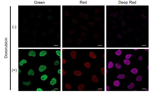
Figure 3. Detection of γH2AX in HeLa cells treated with doxorubicin
| Green | Ex/Em = 488 nm/ 500–550 nm |
| Red | Ex/Em = 561 nm/ 570–620 nm |
| Deep Red | Ex/Em = 640 nm/ 650–700 nm |
| Scale bars | 20 μm |
G265_G266_G267: DNA Damage Detection Kit - γH2AX - Green / Red / Deep Red
Revised Apr., 17, 2023