 Hidden sections will not be printed.
General Information
Exosomes, a form of secreted extracellular vesicles (EVs), contain various proteins and nucleic acids. Consequently, exosomes can have various effects on recipient cells.1) In recent years, numerous studies have reported the involvement of exosomes in processes such as cancer metastasis and malignant progression, therapeutic applications using mesenchymal stem cell-derived exosomes, and diagnostic approaches based on exosomal cargo analysis. Exosomes have accordingly become an important tool in a wide range of biomedical research fields.
ExoIsolator II DirectSpin is an exosome isolation kit designed to achieve a higher recovery rate from cell supernatants than conventional ultracentrifugation (UC). This product has a pre-installed filter in the cup, on which exosomes are trapped by simply adding the cell supernatant to the cup and centrifuging it. This method enables exosome collection without any complex handling procedures.
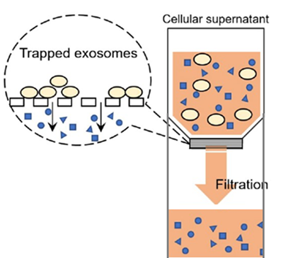 |
| Figure 1. Principle of exosomes purification |
Kit Contents
| 3 tests: | ExoIsolatorⅡ DirectSpin | x3 |
| 10 tests: | ExoIsolatorⅡ DirectSpin | x10 |
| 30 tests: | ExoIsolatorⅡ DirectSpin | x30 |
Storage Condition
Store at room temperature
Required Equipment and Materials
- The 0.22 μm filters
- Used for pre-filtration of the cell supernatant to remove cellular fragments and cell debris.
- Micropipettes
- 50 ml conical tube
- Phosphate-buffered saline (PBS)
- Centrifuge
- Compatible with fixed-angle rotors for 50 ml conical tubes.
- A model with temperature control is recommended.
Precaution
- This product is intended for single use only. Do not reuse the filter cup after washing.
- The membrane filter should be handled with care owing to its thin and fragile structure.
- The recommended sample volume is 1–10 ml. The time required for purification depends on the sample type.
- When using a sterile filter with a pore size larger than 0.22 μm (e.g., 0.45 μm) for pre-filtration, the subsequent centrifugation step may take longer because of clogging.
Preparation
-
Step 1
Prepare the cell supernatant for the experiment.
-
Step 2
Pass the cell supernatant through a 0.22 μm sterile filter to prepare the pre-filtered sample.
General Protocol
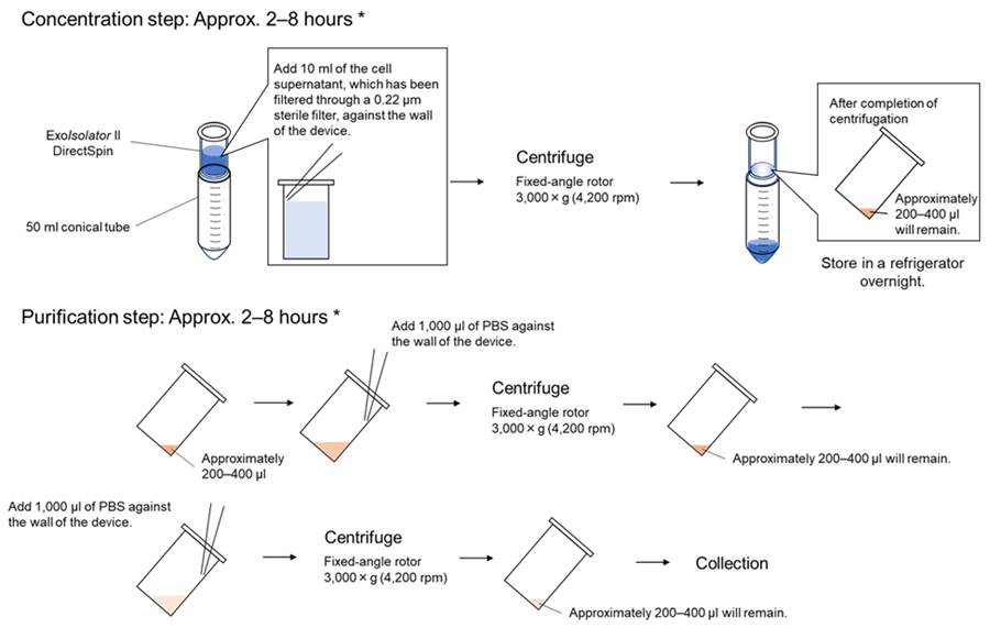
- Set the ExoIsolator II DirectSpin (purification device) into a 50-ml conical tube, add 1–10 ml of the sample solution along the inner wall of the device, and gently close the cap.
- The sample should not be added directly onto the membrane filter. Excessive tightening of the cap may cause damage. Tighten the cap lightly until it stops.
- Centrifuge at 3,000 × g (4,200 rpm) at 4 ℃ for approximately 2–8 h (Concentration step).
- The centrifugation time depends on the sample. When using a new sample, check the liquid volume every 1 h and centrifuge until the residual volume reaches approximately 200–400 μl. If only one purification device is used, prepare a counterbalance tube weighing the same as the 50-ml conical tube containing the sample (adjust the weight by adding water) and centrifuge symmetrically.
- After completion, add 1 ml of PBS against the inner wall of the device.
- If you perform this step on the following day, store the entire 50 ml conical tube containing the sample in a refrigerator (4 ℃) overnight.
- Centrifuge at 3,000 × g (4,200 rpm) at 4 ℃ for approximately 0.5–2 h (first purification step).
- The centrifugation time depends on the sample. Centrifuge until the residual volume reaches approximately 200–400 μl.
- After completion, add 1 ml of PBS along the inner wall of the device again.
- Centrifuge at 3,000 × g (4,200 rpm) at 4 ℃ for approximately 0.5–2 h (second purification step).
- The centrifugation time depends on the sample. Centrifuge until the residual volume reaches approximately 200–400 μl.
- After completion, using a micropipette, gently wash the filter surface with the remaining solution in the device to resuspend the trapped exosomes (approximately 20 times).
- Be careful not to let the pipette tip touch the filter directly.
- Transfer the exosome-suspended solution (approximately 200–400 μl) into a clean 1.5-ml microcentrifuge tube.
- Confirm the volume of the collected exosome solution.
- After adding fresh PBS onto the filter surface, perform flushing again to resuspend the exosomes on the filter (approximately 20 times).
- Add PBS to a final volume of 500 μl, using the solution volume confirmed in step 8 (e.g., if 300 μl was collected in step 8, add 200 μl of PBS).
- Collect the entire volume of the suspended exosome solution and transfer it to the same 1.5-ml microcentrifuge tube used in step 8 to obtain a total of 500 μl of exosome suspension.
 |
| *The centrifugation time varies depending on the cell supernatant sample |
| Operation Figures |
Experimental example
Isolation of exosomes from HEK293S cell supernatant using ExoIsolatorⅡ DirectSpin
- HEK293S cells suspended in 30 ml of serum-free medium at 5 × 10⁵ cells/ml were placed in a 125 ml baffled Erlenmeyer flask. The cells were shaken at 155 rpm at 37 ℃ for 2 days in a 5% CO2 incubator.
- After centrifuging at 1,500 rpm for 5 min, the cell supernatant was transferred to a 50 ml conical tube and stored at 4 ℃ (cell supernatant ①).
- The collected cells were resuspended in 30 ml of fresh serum-free medium and cultured for another 2 days under the same conditions.
- After centrifuging at 1,500 rpm for 5 min, the cell supernatant was transferred to a new 50 ml conical tube and stored at 4 ℃ (cell supernatant ②).
- The cell supernatants collected in steps 2 and 4 were mixed and passed through a 0.22 μm sterile filter to prepare the pre-filtered sample.
- The ExoIsolator II DirectSpin was set into a 50 ml conical tube, 10 ml of the pre-filtered sample was added, and the cap was closed gently.
- Centrifugation was performed at 3,000 × g (4,200 rpm) at 4 ℃ for 4.5 h (approximately 300 μl residual volume).
- After completion, the entire 50 ml conical tube containing the sample was stored in a refrigerator (4 ℃) overnight.
- On the following day, 1 ml of PBS was added against the inner wall of the device, and centrifugation was performed at 3,000 × g (4,200 rpm) at 4 ℃ for 2 h (approximately 300 μl residual volume).
- An additional 1 ml of PBS was added against the inner wall of the device, and centrifugation was performed at 3,000 × g (4,200 rpm) at 4 ℃ for 2 h (approximately 300 μl residual volume).
- The remaining solution in the device was flushed against the filter surface to resuspend the exosomes (approximately 20 times), and 300 μl of the exosome-suspended solution was transferred to a 1.5 ml microcentrifuge tube.
- After adding 200 μl of fresh PBS onto the filter surface, flushing was performed again to resuspend the exosomes on the filter (approximately 20 times).
- The entire volume of the exosome-suspended solution was collected and transferred to the same 1.5 ml microcentrifuge tube used in step 11 to obtain a total of 500 μl of exosome suspension.
- The obtained exosomes were analyzed by nanoparticle tracking analysis (NTA) and western blotting.
NTA
The particle number of purified exosomes was measured using a NanoSight NS300 (Quantum Design, camera level, 13, detection threshold, 5).
Western blotting
- Purified exosomes were mixed with the sample buffer without dithiothreitol and boiled at 95 ℃ for 5 min.
- Samples were separated by SDS-PAGE (SuperSep Ace, 10–20%, 13-well, Wako) and transferred to PVDF membranes (Trans-Blot Turbo Transfer System Transfer Pack, Bio-Rad).
- The following antibodies were used for the detection of CD63 and CD9.
Primary antibody Secondary antibody CD63 Monoclonal mouse anti-CD63 antibody
(diluted 1:1000, cat. no. MEX002-3, MBL)Polyclonal goat anti-mouse antibody conjugated with
HRP (diluted 1:2000, cat. no. ab205719, abcam)CD9 Monoclonal rabbit anti-CD9 antibody
(diluted 1:1000, cat. no. ab236630, abcam)Polyclonal goat anti-rabbit antibody conjugated with
HRP (diluted 1:2000, cat. no. ab97051, abcam) - The membranes were treated with SuperSignal West Pico PLUS Chemiluminescent Substrate (Thermo Fisher Scientific).
- The protein bands were detected using a Syngene Pxi (Syngene).
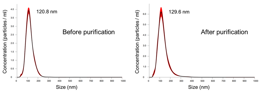 |
|
Figure 2. Particle size distribution of collected exosomes |
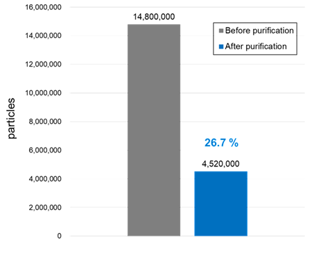 |
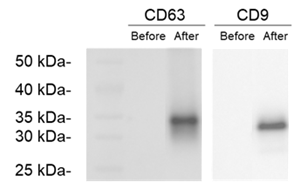 |
| Figure 3. Total particle numbers and recovery rate before and after purification | Figure 4. Comparison of exosome marker expression levels before and after purification |
References
- Kalluri, R.; LeBleu, V. S. The biology, function, and biomedical applications of exosomes. Science. 2020,367(6478).
Frequently Asked Questions / Reference


EX12: ExoIsolator II DirectSpin
Revised Apr., 29, 2026