 Hidden sections will not be printed.
General Information
Endocytosis is a cellular process that involves macromolecules being taken up through a plasma membrane-derived vesicle called an endosome. The endocytic pathway contributes to the maintenance of intracellular homeostasis by bringing in various nutrients to the cell and transporting unwanted components to the lysosome, which acts as a waste disposal system. Recent findings reveal that disruption of endocytosis is related to certain neurodegenerative disorders and immune diseases. Consequently, investigation of the endocytic pathway is attracting considerable interest in the scientific community.
Fluorescent dextran conjugates, internalized via endocytosis, are widely used for monitoring the endocytic pathway in living cells. However, the dextran size affects both internalization and dynamics in the endocytic pathway, suggesting that care should be taken when using dextran conjugates. Therefore, a reliable tool for monitoring the intracellular dynamics of endocytosis is needed.
ECGreen is a small molecule fluorescent probe that is not membrane permeable. ECGreen is internalized via endocytosis and its fluorescence intensity is enhanced as the acidity increases. Unlike conventional dextran conjugates, ECGreen allows direct observation of the membrane of an internalized vesicle, making it suitable for live-cell imaging of endocytosis.
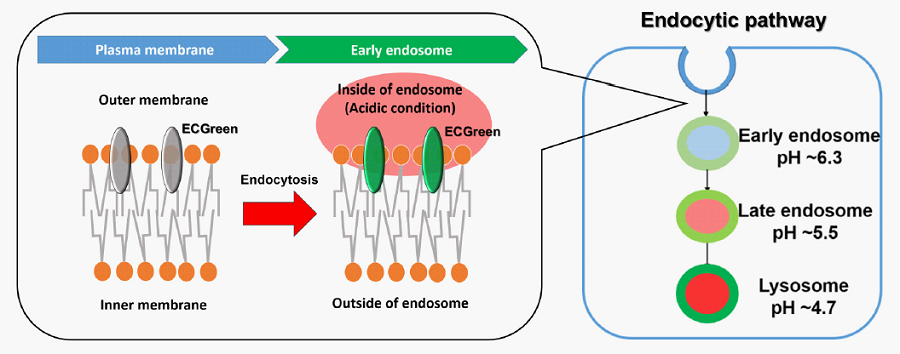
Figure 1. The detection mechanism of endocytosis
Fluorescent Property
Excitation and emission spectra of ECGreen
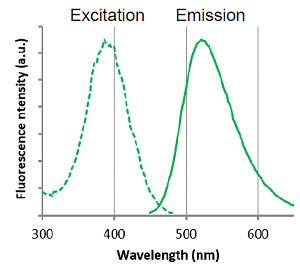 |
λex: 386 nm λem: 522 nm Ex: 350 - 410 nm Em: 500 - 560 nm |
Content
| E296 ECGreen-Endocytosis Detection | 40 μl x 1 (corresponds to 20 dishes (35 mm)) |
Storage Condition
Store at -20 oC
Required Equipment and Materials
- Growth medium
- HBSS or PBS
- Micropipettes
- Microtubes
Preparation of Solution
Preparation of ECGreen working solution
- Dilute the ECGreen solution 1000–5000 times with growth medium to prepare the ECGreen working solution.
- The final concentration of ECGreen should be optimized depending on the cell line.
- The ECGreen working solution should be used on the same day of dilution.
General Protocol
- Seed cells in a dish and culture them overnight at 37 oC in an incubator equilibrated with 95% air and 5% CO2.
- Discard the culture medium and wash the cells once with HBSS, PBS or growth medium.
- Add the ECGreen working solution to the dish containing the cells and incubate them for 10 minutes – 24 hours at 37 oC in an incubator equilibrated with 95% air and 5% CO2.
- Discard the supernatant and wash the cells twice with HBSS, PBS or growth medium.
- Add growth medium to the dish, then observe the cells under a fluorescence microscope.
Usage Example
Observation of endocytosis inhibition by low temperature incubation in HeLa cells
- HeLa cells in MEM (containing 10% fetal bovine serum) were seeded (1.0 × 10⁴ cells/well) on a μ-slide 8 well plate (ibidi) and cultured overnight at 37 oC in an incubator equilibrated with 95% air and 5% CO2.
- The cells were incubated at 4 oC for 30 minutes before staining to test for endocytosis inhibition.
- After washing once with PBS, 200 μl of ECGreen working solution (1000 times dilution) was added to the plate, and the cells were incubated at 37 oC or 4 oC for 30 minutes.
- The cells were washed twice with PBS.
- MEM was added to the plate, and the cells were observed under a confocal fluorescence microscope.
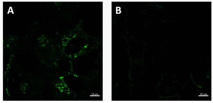
Figure 2. The effect of low temperature incubation on endocytosis
A : Normal condition (37oC incubation), B : Endocytosis inhibition (4oC incubation)
ECGreen filter sets: 405 nm (Ex), 500 – 560 nm (Em)
Frequently Asked Questions / Reference


E296: ECGreen-Endocytosis Detection
Revised Sep., 12, 2023