 Hidden sections will not be printed.
General Information
Autophagy is a process of ordered degradation of dysfunctional cytoplasmic components such proteins and organelles. In this process, an isolation membrane forms in the cytosol, composed of double membrane which gradually expands to enfold aggregated proteins and damaged organelles. The membrane closes to form autophagosomes, which fuse with lysosomes forming autolysosomes. These autolysosomes create acidic compartments, and the contents are decomposed by digestive lysosomal enzymes. Autophagy is thought to be related to aging and neurodegenerative diseases such as Parkinson’s disease, and so there is demand for a simple method of autophagy detection that could be used for drug screening.
The small fluorescent molecule DAPRed is used to be detect autophagosomes and autolysosomes. The mechanism has been suggested to be that the dye is incorporated into the autophagosome during double-membrane formation via structural features, and then emits fluorescence under hydrophobic conditions. The utility of DAPRed is conferred by its molecular properties: it is permeable to cells, has no requirement for transfection, and enables live cell imaging with fluorescence microscopy. For monitoring autolysosomes, DALGreen [D675] is recommend because it enables the detection of phagosome-lysosome fusion1).
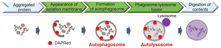
Fig. 1 The detection of autophagy with DAPRed
Content
| DAPRed - Autophagy Detection | 5 nmol x 1 |
Storage Condition
Store at 0–5℃ and protect from light
Required Equipment and Materials
- Dimethyl sulfoxide (DMSO)
- Culture medium
- Hank’s Balanced Salt Solution (HBSS) or serum-free medium
- Micropipettes
Preparation of Solutions
Preparation of 0.1 mmol/L DAPRed DMSO stock solution
Add 50 μLof DMSO to the provided tube containing DAPRed (5 nmol), and dissolve by pipetting.
- Store the reconstituted DAPRed DMSO stock solution at -20℃ and protect from light until use. The solution is stable at -20℃ for 1 month.
Preparation of DAPRed working solution
Dilute the 0.1 mmol/L DAPRed DMSO stock solution with culture medium to prepare 0.1 μmol/L DAPRed working solution.
- Please note that concentration of DAPRed working solution may differ depending on cell line. Therefore please optimize the final concentration of DAPRed working solution for each cell lines.
General Protocol
Autophagy detection
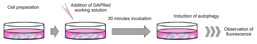
- Prepare cells in a dish.
- Remove the supernatant by aspiration and discard,then wash the cells with culture medium.
- Add an appropriate volume of DAPRed working solution and incubate at 37℃ for 30 minutes.
- Remove the supernatant by aspiration, then wash the cells twice with culture medium.
- Add medium containing the autophagy-inducing agent and incubate at 37℃.
- Please optimize the incubation time according to the conditions of autophagy-induction.
- Observe the cells under a fluorescence microscope.
| Recommended filter | Excitation (nm) | Emission (nm) |
| 500–560 | 690–750 |
Experimental Example
Observation Under the Confocal Fluorescence Microscope
HeLa cells were seeded on CELLview 10 well slide and cultured at 37℃ overnight in a 5% CO2 incubator. Cells were washed with culture medium (Thermo Fisher Scientific, Minimum Essential Media (MEM) supplemented with 10% (v/v) fetal bovine serum, 2 mM L-glutamine, 1% nonessential amino acids, 100 units/mL penicillin, and 100 μg/mL streptomycin), then 100 μL of 0.1 μmol/L DAPRed working solution was added and the cells were incubated at 37℃ for 30 minutes. The culture medium was removed by aspiration and the cells were washed twice with the culture medium, then culture medium or amino acid-free medium (FUJIFILM Wako Pure Chemical Industries, Ltd., Catalogue code: 048-33575) was added to the well. After 6 hours of incubation, the supernatant was removed by aspiration and 100 μL of serum-free medium was added. The cells were then observed by confocal fluorescence microscopy (Fig 2).
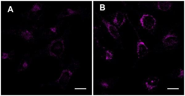 Figure 2 Representive confocal microscope images of HeLa cells stained with DAPRed. |
Excitation wavelength : 561 nm Emission filter : 600–700 nm Scal bar : 20 μm |
Images show micrographs of cells cultured with (A) MEM or with (B) amino acid-free medium. Fluorescence images were taken using a confocal microscope.
Supplemental Information
Excitation and emission spectra of DAPRed
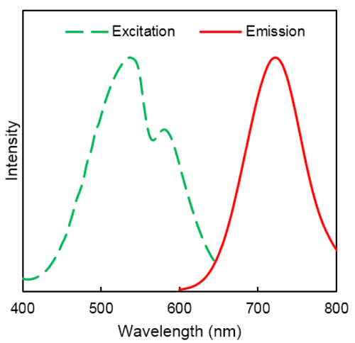 |
λex : 530 nm λem : 720 nm |
Reference
- H. Iwashita, H. T. Sakurai, N. Nagahora, M. Ishiyama, K. Shioji, K. Sasamoto, K. Okuma, S. Shimizu,and Y. Ueno, ‘’Small fluorescent molecules for monitoring autophagic flux’’, FEBS Lett., 2018, 592, 559-567.
Frequently Asked Questions / Reference


D677: DAPRed - Autophagy Detection
Revised May., 22, 2023