 Hidden sections will not be printed.
General Information
Apoptosis is one of the programmed cell death, that plays an important role in maintaining the homeostasis and developmental processes in both plants and animals. Any abnormal cells during the cytogenesis are eliminated by apoptosis. For instance, tumor growth from cancer cells occurred in vivo is inhibited by induction of apoptosis.
However, apoptosis is not induced when the error occurs in tumor suppressor gene p53. Thus, the growth of cancer cells has been found to proceed. Apoptotic cells can be identified based on the alteration of cellular morphology as well as the alternation of biomedical changes.
As of today, various indicators have been established such as caspase activity variation, DNA fragmentation and phosphatidylserine transition on the cell surface-chromosomal. Annexin V stained cells are used to indicate cell membrane changes that occur in the early stage of apoptosis. Once apoptosis is initiated, the phosphatidylserine presents in the inner cell membrane migrates through the cell membrane of the lipid bilayer.
Annexin V specifically binds to phosphatidylserine in the presence of protein-dependent Ca ion. By using fluorescent-labeled Annexin V, the apoptotic cells can be identified by flow cytometry or fluorescence microscopy.
Contents of this kit
| Annexin V, FITC Conjugate | 50 assays x 1 |
| PI Solution | 50 assays x 1 |
| Annexin V Binding Buffer | 50 assays x 1 |
- One assay corresponds to the assay with cell concentration of 1 x 10⁶ cells / mL.
Storage
Do not freeze. Store the kit at 0-5 °C and protect it from long exposure to light.
Precaution
Both FITC-labeled Annexin V and PI are light sensitive. All staining procedures must be performed without direct exposure to intense light.
Required Equipments and Materials
- Flow cytometer or fluorescence microscope
- Approximate fluorescence maximum excitation/emission: Annexin V, FITC: 494 nm / 518 nm; PI: 535 nm / 617 nm
- Adjustable pipettes
- 6-, 12-, 24-, or 96-well plates for cell culturing
- Tubes
- Phosphate buffered saline (PBS)
- Deionized water
- Samples and Inducing agent
Preparation of Reagent solution
10-fold diluted Annexin V Binding Solution
- Dilute Annexin V Binding Buffer by 10-fold with distilled water.
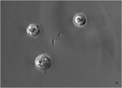
A: Bright Field
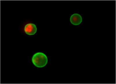
B: Fluorescent Image
Figure 1 Fluorescent imaging of apoptosis induced cells Jurkat cells were apoptosis induced with staurosporine with its concentration of 1 μg/mL at 37 °C for 3.5 hours and then observed under a fluorescent microscope.
General protocol for Suspension Cell
- Centrifuge the cell suspension at 1,000 rpm for 3 minutes and remove supernatant.
- Add PBS for wash cells and centrifuge at 1,000 rpm for 3 minutes, remove supernatant.
Repeat this step one more time. - Add 10-fold diluted Annexin V Binding Solution to make final cell concentration of 1 x 10⁶ cells / mL.
- Transfer 100 μL of cell suspension prepared at step 3 to a new tube.
- Add 5 μL of Annexin V, FITC Conjugate, then 5 μL of PI Solution to the cell suspension.
- Incubate 15 minutes at room temperature with protect from light.
- Add 400 μL of 10-fold diluted Annexin V Binding Solution.
- Apply the solution prepared in step 7 to flow cytometric assay or microscopic assay.
General protocol for Adherent Cells
- Discard supernatant on the petri dish or plate.
- Add PBS for wash cells and discard supernatant. Repeat this step one more time.
- Detach the cells with Trypsin-EDTA.
- Add appropriate volume of culture medium or PBS and transfer the cell suspension to a tube.
- Centrifuge at 1,000 rpm for 3 minutes. Remove supernatant.
- Add PBS for wash and centrifuge at 1,000 rpm for 3 minutes, remove supernatant.
Repeat this step one more time. - Add 10-fold diluted Annexin V Binding Solution to make final cell concentration of 1 x 10⁶ cells / mL.
- Transfer 100 μL of cell suspension prepared at step 7 to a new tube.
- Add 5 μL of Annexin V, FITC Conjugate, then 5 μL of PI Solution to the cell suspension.
- Incubate 15 minutes at room temperature with protection from light.
- Add 400 μL of 10-fold diluted Annexin V Binding Solution.
- Apply this solution to flow cytometric assay or microscopic assay.
- Although adherent cells are not frequently used for Annexin V, FITC flow cytometric analyses because of avoiding the specific cell membrane damage from a cell detachment process, Casiola-Rosen et al. and van Engelend et al. have reported methods on utilizing Annexin V for flow cytometry with adherent cell types.
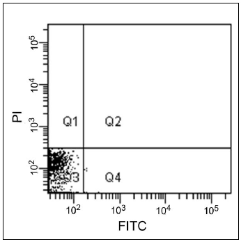
A: Control ( non-treated cells )
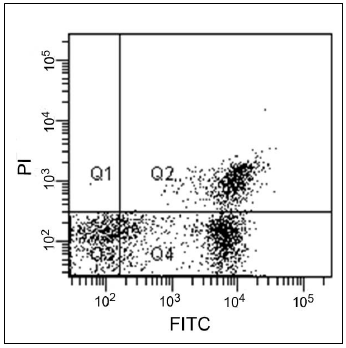
B: Apoptosis induced cells
Figure 2 Flow cytometric analysis of apoptosis induced cells.
Jurkat cells were apoptosis induced with staurosporine with its concentration of 1 μg/mL at 37 °C for 3.5 hours and then analyzed with a flow cytometer.
References
- Casciola-Rosen L, Rosen A, Petri M, Schlissel M, Proc Natl Acad Sci USA, 1996, 93(4), 1624.
- van Engeland M, Ramaekers FC, Schutte B, Reutelingsperger CP, Cytometry, 1996, 24(2), 131.
Frequently Asked Questions / Reference


AD10: Annexin V, FITC Apoptosis Detection Kit
Revised Nov., 15, 2023