

|
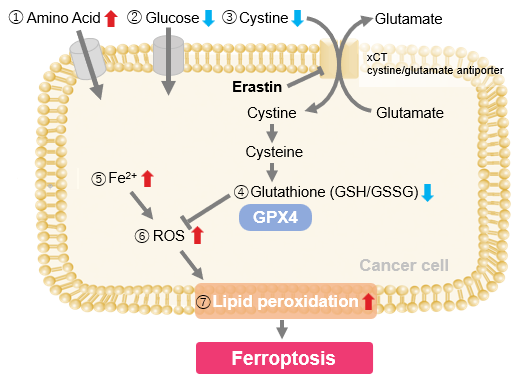
In recent cancer research, ferroptosis, a type of regulated cell death involving iron-dependent lipid peroxidation, has shown promise in cancer treatment. Here are some of the papers that have identified mechanisms of ferroptosis resistance in cancer cells and suggested that they may be therapeutic targets. Ferroptosis, a type of regulated cell death involving iron-dependent lipid peroxidation, has shown promise in cancer treatment. It provides an alternative pathway to eliminate cancer cells that are resistant to traditional therapies such as apoptosis. By targeting ferroptosis, researchers can exploit metabolic weaknesses in cancer cells, particularly when antioxidant defenses are disrupted. This approach is being explored to improve the efficacy of existing treatments and potentially overcome drug resistance in several types of cancer. |
||||||||||||||||||||
|
7-Dehydrocholesterol is an endogenous suppressor of ferroptosis |
Tumor suppressor Par-4 activates autophagy-dependent ferroptosis |
BRCA1-Mediated Dual Regulation of Ferroptosis Exposes a Vulnerability to GPX4 and PARP Co-Inhibition in BRCA1-Deficient Cancers |
||||||||||||||||||
|
Point of Interest - On the other hand, 7-DHC accumulation confers a prosurvival function in cancer cells. - 7-DHC effectively protects (phospho)lipids from autoxidation and subsequent fragmentation due to its superior reactivity with peroxyl radicals. - Accumulation of 7-DHC induces a shift to a ferroptosis-resistant state in tumors. |
Point of Interest - Upregulation of Par-4 promotes NCOA4-dependent ferritinophagy, a selective autophagy process that degrades ferritin, increasing free iron release, lipid peroxidation, and ROS production. - Par-4 inhibition blocks ferroptosis-driven tumor suppression, highlighting its potential for cancer therapy. |
Point of Interest - Combined PARP and GPX4 inhibition in BRCA1-deficient cancers promotes ferroptosis, providing a strategy to overcome PARP inhibitor resistance. - Reduced GPX4 expression and NCOA4-induced ferritinophagy in BRCA1-mutant cancers increases sensitivity to co-treatment with PARP and GPX4 inhibitors. |
||||||||||||||||||
| Related Techniques | ||||||||||||||||||||
| Intracellular / mitochondrial ferrous ion (Fe2+) detection | FerroOrange(intracellular), Mito-FerroGreen(mitochondrial) | |||||||||||||||||||
| Lipid peroxidation detection | Liperfluo(intracellular), MitoPeDPP(mitochondrial) | |||||||||||||||||||
| First-time autophagy research | Autophagic Flux Assay Kit | |||||||||||||||||||
| Total ROS detection | Highly sensitive DCFH-DA or Photo-oxidation Resistant DCFH-DA | |||||||||||||||||||
| Mitochondrial superoxide detection | MitoBright ROS Deep Red - Mitochondrial Superoxide Detection | |||||||||||||||||||
| Cellular senescence detection | SPiDER-βGal for live-cell imaging or flow cytometry / microplate reader / tissue samples | |||||||||||||||||||
| Glutathione Quantification | GSSG/GSH Quantification Kit | |||||||||||||||||||
| Cystine Uptake detection | Cystine Uptake Assay Kit | |||||||||||||||||||
| Glycolysis/Oxidative phosphorylation Assay | Glycolysis/OXPHOS Assay Kit | |||||||||||||||||||
| Mitochondrial membrane potential detection | JC-1 MitoMP Detection Kit, MT-1 MitoMP Detection Kit | |||||||||||||||||||
| Apoptosis detection in multiple samples | Annexin V Apoptosis Plate Assay Kit | |||||||||||||||||||
| Cell proliferation/ cytotoxicity assay | Cell Counting Kit-8 and Cytotoxicity LDH Assay Kit-WST | |||||||||||||||||||
| Related Applications | ||||||||||||||||||||
Erastin-Induced Ferroptosis: Evaluating Intracellular Uptake and Redox Balance
|
||||||||||||||||||||
Exploring Ferroptosis Resistance Mechanisms for Cancer Treatment

Related Selection Guide & Science Note


