


Glucose Uptake Plate Assay Kit

Glucose uptake capacity microplate assay
- Glucose uptake capacity can be easily measured with a microplate reader
- Non-wash type that does not require any washing
- Simple operation: only requires adding reagents
-
Product codeUP08 Glucose Uptake Plate Assay Kit
| Unit size | Price | Item Code |
|---|---|---|
| 100 tests | Find your distributors | UP08-10 |
| 500 tests | Find your distributors | UP08-12 |
[Note]
For measurement, please use a clear bottom 96-well black plate and a plate reader capable of bottom excitation and fluorescence measurement.
| 100 tests | ・Glucose Uptake Probe-Green ・Quenching Buffer |
×1 11 ml×1 |
|---|---|---|
| 500 tests | ・Glucose Uptake Probe-Green ・Quenching Buffer |
×5 11 ml×5 |


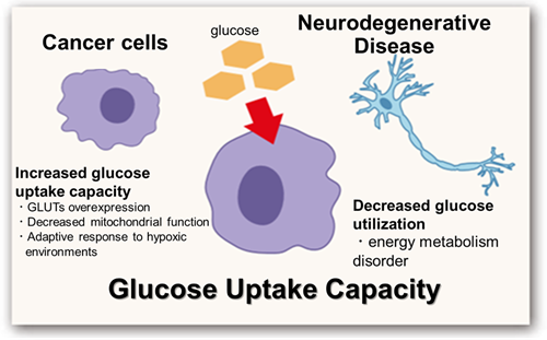
The relationship between glucose uptake and disease
Metabolism, the process by which cells absorb nutrients and produce energy, has recently been found to control various cellular functions, including gene expression.
In cancer cells, glucose uptake is significantly increased. This metabolic characteristic, known as the Warburg effect, is regulated by several mechanisms, including the overexpression of glucose transporters (GLUTs), impaired mitochondrial function, and adaptation to hypoxic environments. These changes are being closely monitored as indicators of cancer malignancy and treatment resistance.
Conversely, impaired glucose utilization and metabolic abnormalities have been shown to be closely related to neurological dysfunction in neurodegenerative diseases such as Alzheimer's disease.
Thus, visualization of glucose metabolism has become an important indicator for understanding disease mechanisms and developing treatments.
Glucose Uptake Assay Products
| Products | Plate Reader | Microscope / FCM |
|---|---|---|
| Glucose Uptake Assay Kit-Blue | ✗ | ✓ |
| Glucose Uptake Assay Kit-Green | ✓ | ✓ |
| Glucose Uptake Plate Assay Kit (Green) | ✓ Best* | ✗ |
| Glucose Uptake Assay Kit-Red | ✓ | ✓ |
*No washing step, minimizing cell stress and detachment while improving reproducibility.
Manual
Technical info
Cells take in various nutrients and produce energy via intracellular metabolism. The metabolism of these nutrients varies depending on the extracellular environment, cell state, and cell type. In recent years, it has become clear that nutrient metabolism plays a role not only in energy production, but also in controlling various cellular functions, including gene expression.
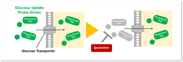
Glucose is a primary nutrient, so understanding its uptake and metabolism is crucial for elucidating cellular functions. This kit includes a fluorescently labeled glucose derivative, Glucose Uptake Probe-Green, which is taken up by cells via glucose transporters. This enables the measurement of cellular glucose uptake capacity through fluorescence measurement using a plate reader. Any Glucose Uptake Probe-Green not taken up by the cells can be quenched using the Quenching Buffer. This eliminates the need to wash the probe, making the assay more convenient with a plate reader.
Breakthrough: Quenching Buffer solves wash step limitations
The product includes a Quenching Buffer, removing the need for washing after reagent addition It addresses the following customer issues:
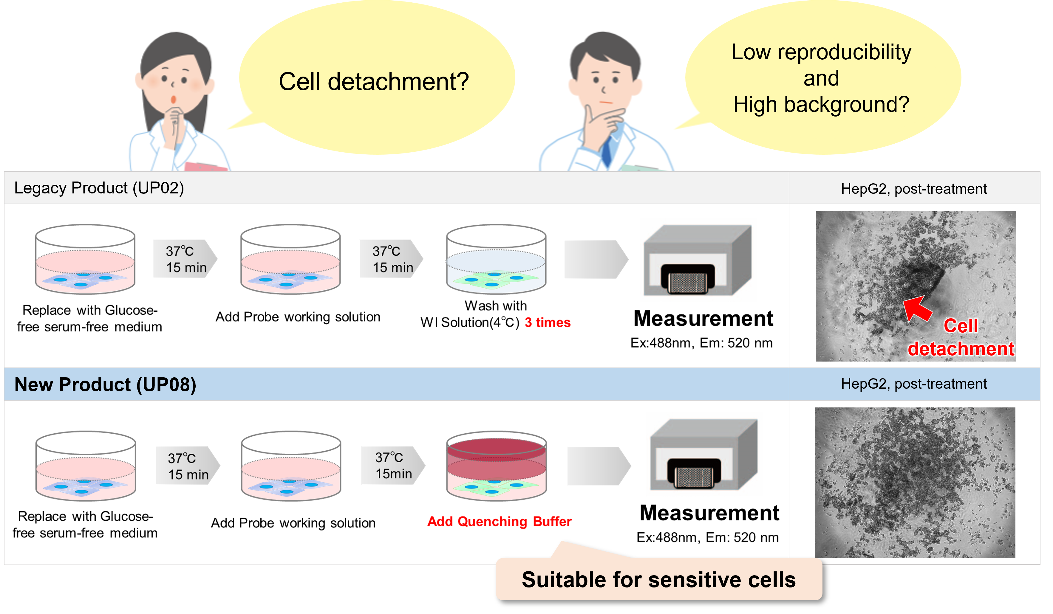
Conventional product:Glucose Uptake Assay Kit-Green (code:UP02)
Product comparison
The comparison table for the Glucose Uptake Assay Kit-Green (product code : UP02) is shown below.
This product is a glucose uptake kit designed for plate assays. It is ideal for those who wish to perform plate assays with minimal effort and avoid cell detachment because it does not require washing of the probe working solution.
Conversely, the Glucose Uptake Assay Kit-Green (Product Code : UP02) is recommended for plate assays, flow cytometry, or imaging evaluations in cell types where detachment due to washing procedures is not a concern.
| DOJINDO LABOLATORIES | ||
|---|---|---|
| Code | UP08 | UP02 |
| Product Name |
This Product |
|
|
Compatible device |
Plate reader |
Fluorescence microscope, Plate reader, |
| Wash Type |
ー |
✓ |
|
Non-Wash Type |
✓ |
ー |
|
Measurement target |
Live cell |
Live cell |
Can be measured even with cell types that are prone to detachment
This product is designed to minimize cell detachment, allowing more consistent measurements.
 |
When using cell types that peel off during medium replacement |
<Protocol>
1. Poly-D-Lysine (PDL) solution (1mg/ml) was diluted 10-fold with PBS(-).
2. The diluted PDL solution (150 µl/well) was added to each well of a 96-well microplate (655090, Greiner GmbH), and the plate was incubated at room temperature for 1 hour.
3. Each well was washed three times with PBS(-) (200 µl/well).
4. HEK293 cells suspension (6.0 × 10⁵ cells/ml, 200 µl/well) in MEM (10% fetal bovine serum、1% penicillin-streptomycin) were seeded in the PDL-coated 96-well microplate and cultured at 37°C overnight in a 5% CO2 incubator.
5. 2-Deoxy-D-glucose (2-DG) solution (900 mmol/l, 22 µl) was added to each well (see Figure 8) (final concentration 100 mmol/l 2-DG), and the cells were incubated at 37°C for 2.5 hours in a 5% CO2 incubator.
6. After removing the supernatant, the cells were washed twice with 200 μl of pre-warmed DMEM (glucose- and serum-free, 37°C).
7. Pre-warmed DMEM (200 μl, glucose- and serum-free, 37°C) was added to each well, and the cells were incubated at 37°C for 15 min in a 5% CO2 incubator.
8. After removing the supernatant, 100 μl of pre-warmed probe working solution in DMEM (glucose- and serum-free, 37°C) was added to each well, and the cells were incubated at 37°C for 30 min in a 5% CO2 incubator.
9. The pre-warmed Quenching Buffer (100 µl, 37°C) was added to each well.
10. The fluorescence intensity was measured using a microplate reader (Infinite M200 PRO, Tecan Trading AG, bottom reading, Ex/Em = 488/520 nm).
※To operate the conventional product (UP02) and take measurements, follow the instructions in the UP02 instruction manual from step 9 onwards.
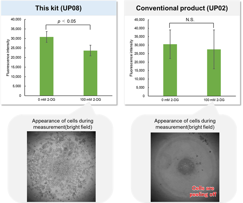
Experimental example: Inhibition of Glucose Uptake by D-Glucose Competition
We evaluated the glucose uptake capacity of A549 cells using this product.
Under high-glucose conditions (25 mmol/l), competitive inhibition occurs between D-glucose and the Glucose Uptake Probe, resulting in reduced fluorescence intensity compared to glucose-free conditions (0 mmol/l).
These results confirm that the uptake of the Glucose Uptake Probe serves as an indicator of glucose uptake capacity.
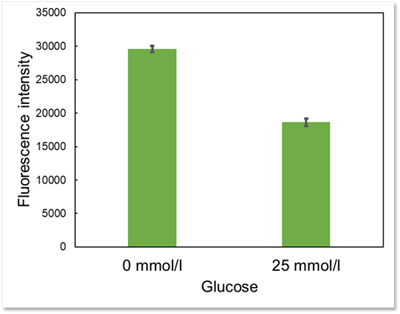
Cells: A549
Detection condition: Ex : 488 nm, Em : 520 nm
Experimental example: Inhibition of Glucose Uptake by Cytochalasin B
Using this kit, we were able to quantify the inhibition of Glucose Uptake in HepG2 cells by the glucose transporter inhibitor Cytochalasin B.
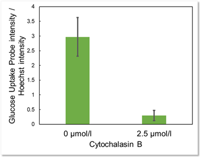
Cells: HepG2
Culture conditions: 2.5 µmol/l Cytochalasin B in DMEM (25 mmol/l Glucose, 10% FBS, 1%P/S), 37℃, overnight
Detection condition: Ex : 488 nm, Em : 520 nm
*Normalized with Hoechst 33342 fluorescence intensity.
Fluorescence property
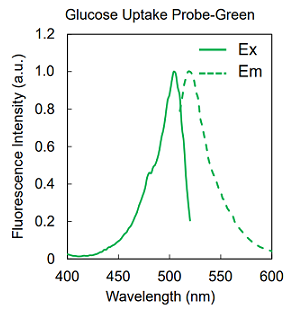
Fig. Excitation and emission spectra of Glucose Uptake Probe-Green
Q & A
-
Q
Could you please explain the difference between this product and the Glucose Uptake Assay Kit-Green (product code: UP02)?
-
A
This product (UP08) is a glucose uptake evaluation kit designed for plate assays. It is ideal for users who wish to simplify their assays or avoid cell detachment because it eliminates the need to wash the probe working solution.
In contrast, the Glucose Uptake Assay Kit-Green (product code: UP02) is suitable for plate assays, flow cytometry, and imaging.
-
Q
Does Glucose Uptake Probe-Green have selectivity for the type of glucose transporter?
-
A
No. Glucose Uptake Probe-Green is believed to be taken up by multiple glucose transporters. However, there are no detailed data on the probe’s selectivity or affinity for specific GLUTs.
-
Q
Is Glucose Uptake Probe-Green degraded and metabolized after uptake into the cell?
-
A
The glucose moiety may be phosphorylated by hexokinase due to its structure, but no further metabolism is expected.
-
Q
After Glucose Uptake Probe-Green is incorporated into cells, can the cells be fixed?
-
A
No. Glucose Uptake Probe-Green leaks out of cells after fixation.
-
Q
Which cell lines have been analysed with this kit?
-
A
We have tested the kit using the following cell lines.
Cell lines Human alveolar basal epithelial adenocarcinoma cells A549 Human hepatoma-derived cells HepG2 Human Embryonic Kidney cells HEK293
-
Q
Please provide the results of experiments altering inhibitor activity or uptake capacity.
-
A
We have performed such experiments in the following cell lines:
Cell lines
Dilution factor of
probe stock solution
staining
time
Inhibitors or experimental
conditions
Human hepatoma-derived cells HepG2 ×500 15分 Cytochalasin B Human Embryonic Kidney cells HEK293 ×500 30分 2-DG
-
Q
Which plate should I use for measurements with the plate reader?
-
A
Since a bottom reading is required for fluorescence detection, use a clear-bottom 96-well black plate.
-
Q
What should I do if the sensitivity of measurement using Glucose Uptake Probe-Green is low?
-
A
Please consider the probe concentration (×250 to ×1,000) and staining time (15 minutes to 1 hour) as initial considerations.
-
Q
Can I save the Probe working solution?
-
A
No, the Probe working solution cannot be stored. The Probe Stock solution can be stored -20 oC for 1 month.
-
Q
Does Glucose Uptake Probe-Green have any cytotoxicity?
-
A
Glucose Uptake Probe-Green has no cytotoxicity within the range of manual use. Using our product Cell Counting Kit-8 (product code: CK04), we measured the cytotoxicity of this probe in A549 cells.
-
Q
Can the Glucose Uptake Assay Kit-Green be used to quantify glucose in the culture medium or in cells?
-
A
No. Please use our product, Glucose Assay Kit-WST (product code: G264).
-
Q
Is it possible to quantify Glucose Uptake Probe-Green taken up into cells?
-
A
No, this kit measures the fluorescence intensity of the probe taken up into the cell as an indication of the glucose uptake capacity of the cell.
-
Q
Can it be used for imaging with a fluorescence microscope or for flow cytometry measurements?
-
A
No. For imaging or flow cytometry of probes internalized into cells, use the Glucose Uptake Assay Kit-Green (product code: UP02).
-
Q
How long is the probe retained in the cells after adding the Quenching Buffer?
-
A
After adding the Quenching Buffer, the probe gradually moves into the extracellular space. To minimize measurement variability, perform the measurement within 10 minutes of adding the Quenching Buffer. However, this time frame may vary by cell type.
-
Q
What should I do if cells detach when switching to glucose-free serum-free medium?
-
A
If this happens, consider using plates coated with poly-D-lysine (PDL), as described in the following procedure.
<Protocol>
1. Poly-D-Lysine (PDL) solution (1mg/ml) was diluted 10-fold with PBS(-).
2. The diluted PDL solution (150 µl/well) was added to each well of a 96-well microplate (655090, Greiner GmbH), and the plate was incubated at room temperature for 1 hour.
3. Each well was washed three times with PBS(-) (200 µl/well).
4. HEK293 cells suspension (6.0 × 10⁵ cells/ml, 200 µl/well) in MEM (10% fetal bovine serum、1% penicillin-streptomycin) were seeded in the PDL-coated 96-well microplate and cultured at 37°C overnight in a 5% CO2 incubator.
5. 2-Deoxy-D-glucose (2-DG) solution (900 mmol/l, 22 µl) was added to each well (see Figure 8) (final conc.100 mmol/l 2-DG), and the cells were incubated at 37°C for 2.5 hours in a 5% CO2 incubator.
6. After removing the supernatant, the cells were washed twice with 200 μl of pre-warmed DMEM (glucose- and serum-free, 37°C).
7. Pre-warmed DMEM (200 μl, glucose- and serum-free, 37°C) was added to each well, and the cells were incubated at 37°C for 15 min in a 5% CO2 incubator.
8. After removing the supernatant, 100 μl of pre-warmed probe working solution in DMEM (glucose- and serum-free, 37°C) was added to each well, and the cells were incubated at 37°C for 30 min in a 5% CO2 incubator.
9. The pre-warmed Quenching Buffer (100 µl, 37°C) was added to each well.
10. The fluorescence intensity was measured using a microplate reader (Infinite M200 PRO, Tecan Trading AG, bottom reading, Ex/Em = 488/520 nm).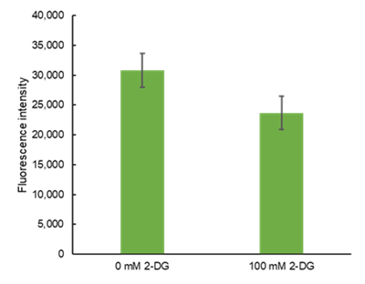
Handling and storage condition
| Store at 0-5°C |


