Senescence Driven by Mitochondrial Alterations [Mar. 5, 2025]
| Cellular senescence is closely linked to mitochondrial dysfunction, which drives aging and disease through metabolic shifts, oxidative stress, and pro-inflammatory signaling. This Science Note shows the latest findings on mitochondrial mechanisms that promote cellular senescence. | ||||||||||||||||||||||||
|
Apoptotic stress causes mtDNA release during senescence and drives the SASP Highlighted technique: When apoptotic stress increases the permeability of the mitochondrial outer membrane, cytochrome c is released into the cytoplasm, making it a key indicator of MOMP. In this study, MOMP is assessed by co-staining the mitochondrial outer membrane protein TOM20 and cytochrome c, followed by high-resolution analysis. |
||||||||||||||||||||||||
|
Mitochondrial fatty acid oxidation drives senescence Highlighted technique: This study assessed senescence using multiple markers, including p16 expression and SA-βgal activity. While other markers changed, γH2AX did not, reinforcing the widely accepted notion that no single marker defines senescence. Thus, multiple indicators are needed to assess senescence. |
||||||||||||||||||||||||
|
Senescent glia link mitochondrial dysfunction and lipid accumulation |
||||||||||||||||||||||||
Related Techniques (click to open/close)
|
||||||||||||||||||||||||
Application Note (click to open/close)
|
||||||||||||||||||||||||
|
|
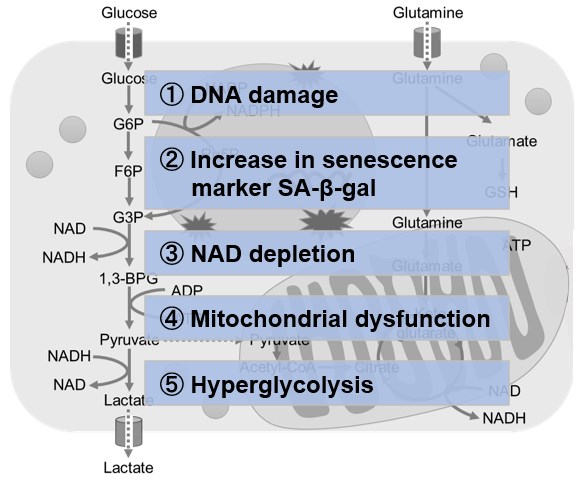
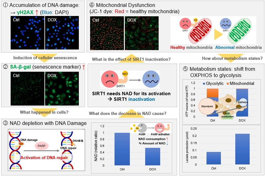
NAD(+) levels decline during the aging process, causing defects in nuclear and mitochondrial functions and resulting in many age-associated pathologies*. Here, we try to redemonstrate this phenomenon in the doxorubicin (DOX)-induced cellular senescence model with a comprehensive analysis of our products. *S. Imai, et al., Trends Cell Biol, 2014, 24, 464-471
|
|
|
|