New function of endocytosis induced and inhibited by membrane proteins [Jun. 26, 2024]

[Jun. 26, 2024] Previous Science Note

Endocytosis induction refers to the initiation of the endocytic process, where specific signals trigger the cell to internalize extracellular substances. This induction often involves membrane proteins that can act as receptors to recognize and bind external ligands, initiating the formation of endocytic vesicles. The interaction between ligands and membrane proteins not only triggers endocytosis, but also ensures that the internalized cargo is selectively processed. Thus, membrane proteins are integral to both the initiation and specificity of endocytosis, influencing cellular uptake and signaling pathways.

Endocytic vesicles act as vehicles for glucose uptake in response to growth factor stimulation
Click here for the original article: Ryouhei Tsutsumi, et. al., Nature Communications, 2024.

Point of Interest

- Platelet-derived growth factor(PDGF) enhances cellular glucose uptake via receptor endocytosis and independently of known mechanisms, resulting in a near doubling of glucose uptake by the cells.

- PDGF receptor (PDGFR) co-endocytoses with subset of glucose transporter 1 (GLUT1/SLC2A1) upon PDGF-stimulation.

- The PDGFR/GLUT1-containing endosomes have multiple glycolytic enzymes and localize to adjacent mitochondria.

- The glucose-loaded endosomes generated by growth factors deliver glucose to the glycolytic machinery in proximity to mitochondria.

Endocytosis blocks the vesicular secretion of exosome marker proteins
Click here for the original article: Yiwei Ai et. al., Science Advances, 2024.

Point of Interest

- Endocytosis inhibition induces plasma membrane accumulation and vesicular secretion of CD63.

- High expression of CD63, CD81 or CD9 inhibits its own endocytosis and induces its plasma membrane accumulation and vesicular secretion.

- Induction of endocytosis inhibits their vesicular secretion and, in the case of CD9 and CD81, causes their destruction in the lysosome.

- Vesicular secretion of exosome marker proteins occurs primarily through an endocytosis-independent pathway.

Cell surface protein aggregation triggers endocytosis to maintain plasma membrane proteostasis
Click here for the original article: David Paul et. al., Nature Communications, 2023.

Point of Interest

- Aggregation of protein ectodomains by cross-linking antibodies ability triggers specific and fast endocytosis of the receptor, independent of clathrin and dynamin.

- Upon aggregation, even canonical clathrin-dependent cargoes are redirected to the aggregation-dependent endocytosis (ADE) pathway.

- ADE is an actin-driven process that morphologically resembles macropinocytosis.

- ADE clears stress-induced receptor aggregates and facilitates their lysosomal degradation to maintain cell surface proteostasis.

Related Techniques

Endocytosis Detection detection
ECGreen-Endocytosis Detection
Exosome Labeling
ExoSparkler Exosome Membrane Labeling Kit-Green / Red / Deep Red
Lysosomal function
Lysosomal Acidic pH Detection Kit -Green/Red and Green/Deep Red
Mitophagy or autophagy detection
Mitophagy Detection KitAutophagic Flux Assay Kit
 
Plasma Membrane Staining
PlasMem Bright Green / Red
Glycolysis/Oxidative phosphorylation Assay
Glycolysis/OXPHOS Assay Kit

Related Applications

Clear visualization of intracellular vesicular trafficking

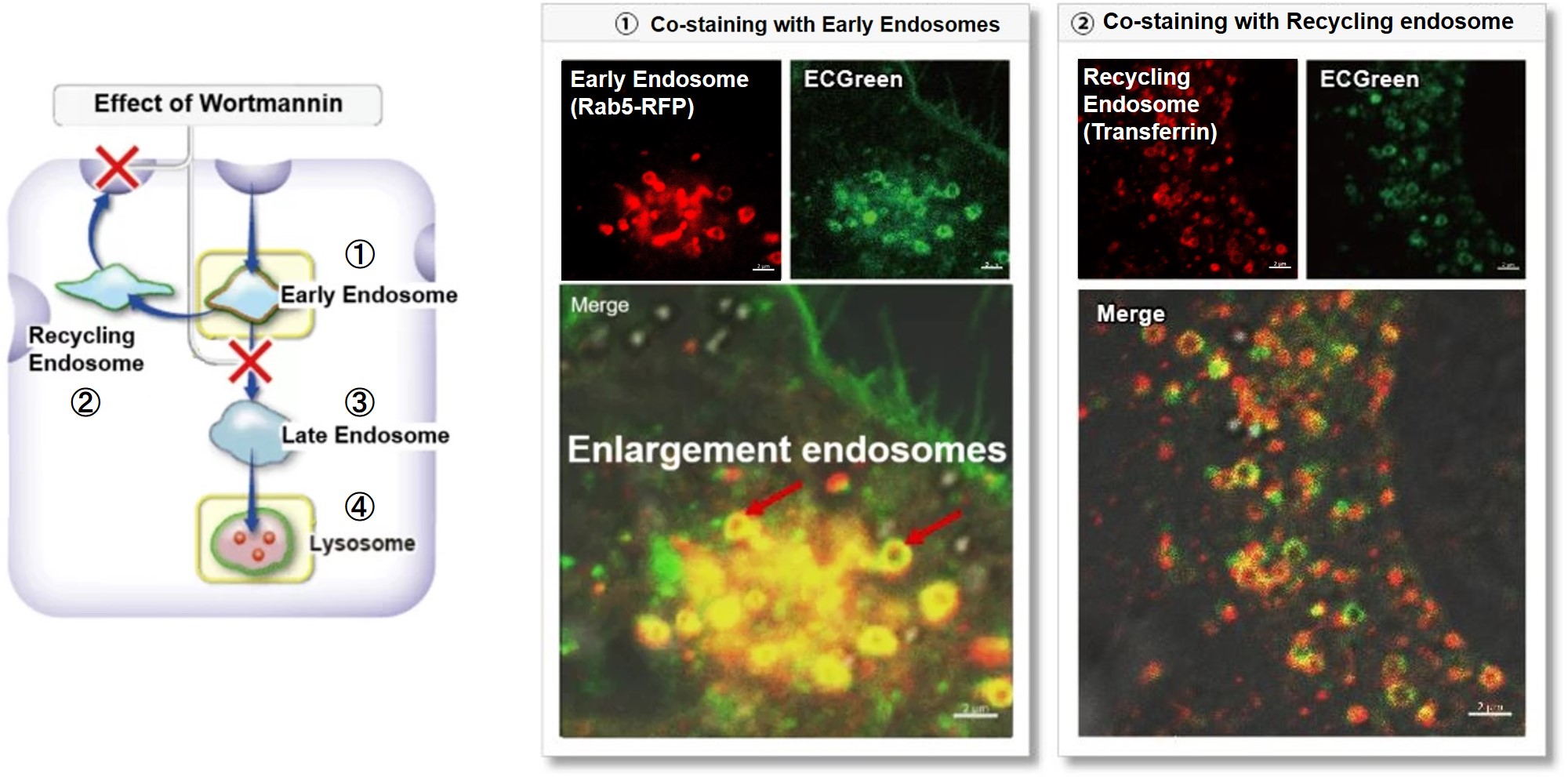
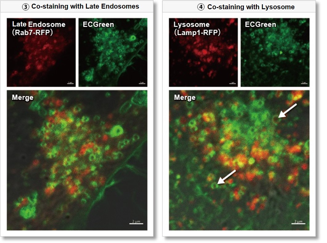
Wortmannin is known to inhibit endosomal recycling and lysosomal translocation, leading to endosomal enlargement.
These changes induced by Wortmannin were confirmed by co-staining with ECGreen (green) and the following indicators.

①Eary endosome: Rab5-RFP (red)
② Recycling endosome: Fluorescent labeled Transferin (red)
③ Late endosome: Rab5-RFP (red)
④ Lysosome: Lamp1-RFP (red)

As a result, it was confirmed that ECGreen (green) co-localizes only with enlarged early endosomes and recycling endosomes (Fig. ① and ②), but not with late endosomes or Lysosomes (Fig. ③ and ④), supporting Wortmannin's effect. ECGreen can visualize changes in the intracellular vesicular trafficking system and endosome shape.

Endosomes (ECGreen, green): Ex. 405 nm / Em. 500 – 560 nm
Early endosomes (Rab5-RFP, red): Ex. 561 nm / Em. 560 – 620 nm
Recycling endosome (Transferrin-Alexa fluor 488 conjugate, red: pseudo-color): Ex. 488 nm / Em. 500 – 550 nm
Late endosomes (Rab7-RFP, red): Ex. 561 nm / Em. 560 – 620 nm
Lysosomes (Lamp1-RFP, red): Ex. 561 nm / Em. 560 – 620 nm
 

[Experimental Procedure]
(1) Prepare HeLa cells in 8 wells of μ-Slide and incubate overnight.
(2) After washing with HBSS, 200 µl of Wortmannin (final concentration: 100 nmol/l) prepared in 10% FBS-containing MEM medium was added.
(3) Incubate at 37°C for 30 minutes
(4) 200 µl of ECGreen (diluted 1,000-fold) prepared in 10% FBS-containing MEM medium without removing the supernatant
(5) Incubate at 37°C for 30 minutes
(6) Wash the cells twice with HBSS and add MEM medium containing 10% FBS.
(7) Observation with a confocal laser microscope

Product Classification

Product Classification