| [Nov. 28, 2023] | Previous Science Note |
|
Scientists have discovered that the mitochondrial translocator protein (TSPO) and hexokinase-2 play key roles in controlling microglial metabolism and phagocytosis. Microglia lacking TSPO resembled dysfunctional microglia observed in aging and Alzheimer's disease, and this could be partially reversed by blocking hexokinase-2 binding to mitochondria. They conclude that targeting mitochondrial hexokinase-2 binding may provide an immunotherapeutic approach to inhibit glycolytic metabolic reprogramming and promote microglial phagocytosis in Alzheimer's disease. |
|
|
Related Techniques |
|
|
|
|
|
|
|
|
|
Related Applications |
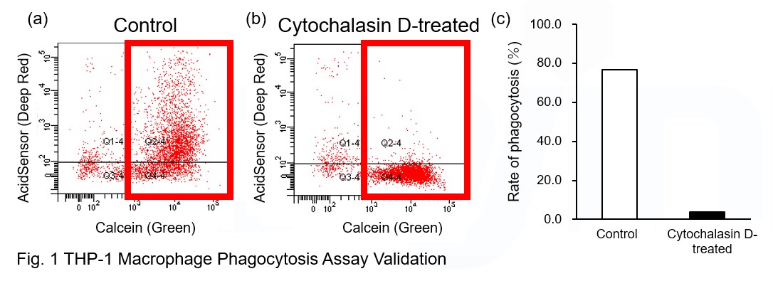
Phagocytosis assay of labeled apoptotic cells in THP-1 cells
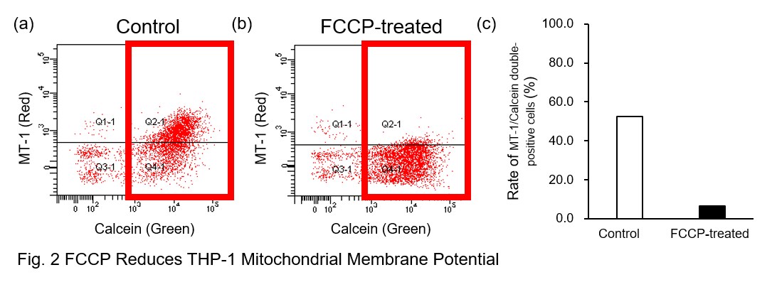
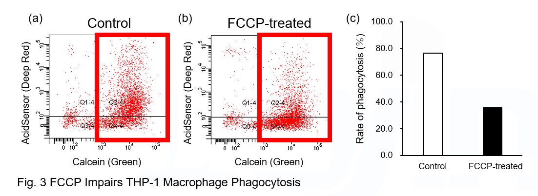
AcidSensor-labeled substances are taken up by cells and their fluorescence increases when they reach acidic organelles such as lysosomes. Taking advantage of this property, we evaluate the phagocytic activity of apoptotic cells by co-culturing AcidSensor-labeled apoptotic cells with Calcein-labeled THP-1 macrophages. As a result, Calcein (Green) / AcidSensor (Deep red) double-positive cells, indicating THP-1 macrophages phagocytosing apoptotic cells, were observed by flow cytometry (Fig. 1a). Furthermore, when the phagocytosis of THP-1 macrophages was inhibited by Cytochalasin D, the percentage of double-positive cells decreased (Fig. 1b and 1c), confirming that the assay system can accurately evaluate phagocytosis. A recent report reveals that inhibition of mitochondrial function induces a switch to glycolysis and reduces phagocytosis in cultured microglia, resident macrophages in the central nervous system*. To replicate this result, phagocytosis assays were performed using mitochondria-inhibited THP-1 macrophages. The results show that FCCP, a potent uncoupler of oxidative phosphorylation in mitochondria, decreases mitochondrial membrane potential (MT-1, Red) of THP-1 macrophages (Fig. 2) and reduces phagocytosis (Fig. 3). *Lauren H. Fairley, et al., PNAS (2023)
|