


Science Note / oxidative stress
Mitochondrial Connections: How Organelle Contact Sites Shape Oxidative Stress and Cell Death
Mitochondria do not act alone. They form dynamic contacts with other organelles, enabling the exchange of metabolites, ions, lipids, and signaling molecules1. These mitochondrial contact sites connect mitochondria with the endoplasmic reticulum (ER)2, lysosomes3, peroxisomes4, 5, nuclei6, and broader networks involving multiple organelles1.
Mitochondria posses extensive redox machinery making them a central source of reactive oxygen species (ROS)7. In excess, mitochondrial-derived ROS can damage DNA, cellular membranes, and promote cell death. Lipid peroxidation is a key form of this oxidative damage, compromising membrane integrity and contributing to cell death.
Recent work has highlighted that stimulus-responsive mitochondrial contacts sites may fuel unique cellular responses during oxidative stress. Below, we discuss two recent studies describing how mitochondrial connections concentrate oxidative stress at specific cellular locations. From localized oxidative damage at the plasma membrane to phospholipid peroxidation at ER–mitochondria contacts, these findings highlight how the location of oxidative stress can shape cell death.
Contact with the Plasma Membrane: Mitochondrial ROS Delivery During Immune Activation and Metabolic Stress
Wang et al.8 developed a model system to assess the coupling of innate immune activation and metabolic disruption. This combination of stimuli induced a unique type of cell death that did not rely on canonical pathways. The authors' description of this cell death pathway revealed localized ROS hotspots that colocalized with mitochondria; following stimulation, mitochondria idled at the plasma membrane, locally accumulated ROS, and damaged the plasma membrane to promote cell death.
This novel form of cell death induced by immune and metabolic stress, driven by prolonged mitochondria-PM contact was named mitoxyperilysis.
Mitoxyperilysis-stimulating conditions were associated with tumor regression in vivo. The authors argue these findings may support therapeutic avenues for cancer treatment that utilize fasting and/or immune activation.
Contact with the ER: Lipid Peroxidation Hubs at the Outset of Ferroptosis
Iron-dependent membrane phospholipid peroxidation is central to the cell death pathway ferroptosis. Lipid peroxidation can be initiated non-enzymatically through reaction of membrane polyunsaturated fatty acids with the hydroxyl radical. Lipid peroxidation therefore represents a damaging outcome of oxidative stress and a defining mechanism of ferroptosis.
While previous studies have implicated lysosomes in early ferroptotic events9, 10, Sassano et al. identified ER–mitochondria contact sites (EMCSs) as initial hotspots of phospholipid peroxidation11.
Live-cell super-resolution microscopy revealed a rapid increase in ER–mitochondria contact sites following ferroptosis stimulation. Shortly after stimulation, lipid peroxidation accumulated at these contact sites and propagated toward mitochondria.
Using genetic tools to narrow, widen, or impair these contacts, the authors showed that closer ER–mitochondria apposition increased sensitivity to ferroptosis. Disrupting connectivity reduced lipid peroxide formation across the cell and increased resistance to ferroptotic death, supporting a role for these contacts in propagating cellular lipid peroxidation requried for ferroptosis progression.
← Previous Science NoteResearch Highlights
Innate immune and metabolic signals induce mitochondria-dependent membrane lysis via mitoxyperiosis
Highlighted Techniques
Using common ROS probes, the authors identified ROS hotspots where probe oxidation was locally higher compared to the whole cell. Using live cell imaging and co-staining of mitochondria it was observed that the ROS hotspots coincided with extended dwelling of mitochondria at the plasma membrane; this dwelling allowed localized ROS accumulation, lipid peroxidation, and plasma membrane rupture.
Endoplasmic reticulum-mitochondria contacts are prime hotspots of phospholipid peroxidation driving ferroptosis
Highlighted Techniques
The authors extensively characterized subcellular localization of lipid peroxidation and lipid peroxides with the probes Liperfluo and BODIPY C11. By simultaneously tracking ER and Mitochondria dynamics, they were able to show rapid, local increases in lipid peroxide accumulation at ER-Mitochondria contact sites following Ferroptosis induction with RSL3.
Application Note
Real experimental data, practical readouts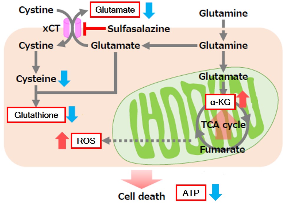
Experimental Data
Application Note I
Sulfasalazine Alters Intracellular Metabolites and Increases ROS in A549 Cells
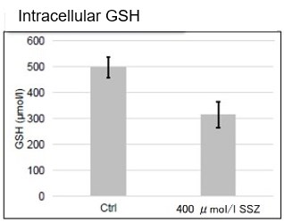
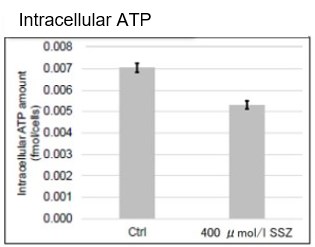
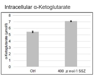
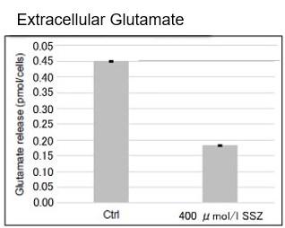
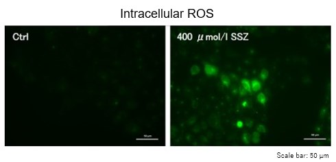
A549 cells were treated with sulfasalazine (SSZ), an inhibitor of the cystine/glutamate transporter xCT. Intracellular GSH, ATP, α-ketoglutarate (α-KG), ROS, and glutamate release were evaluated.
Results
- Intracellular GSH and ATP decreased.
- Glutamate release decreased.
- Intracellular α-KG and ROS increased.
- Cells
- A549, 1 × 10⁶ cells.
- Treatment
- 400 μmol/l sulfasalazine, 48 hours.
- Readouts
- GSH, ATP, α-KG, glutamate release, and intracellular total ROS.
Products in Use
- ① Intracellular Glutathione
GSSG/GSH Quantification Kit - ② Intracellular ATP
ATP Assay Kit-Luminescence - ③ Intracellular α-Ketoglutarate
α-Ketoglutarate Assay Kit-Fluorometric - ④ Glutamate Release
Glutamate Assay Kit-WST - ⑤ Intracellular Total ROS
ROS Assay Kit -Highly Sensitive DCFH-DA-
Reference
Shogo Okazaki et al., Glutaminolysis-related genes determine sensitivity to xCT-targeted therapy in head and neck squamous cell carcinoma. Cancer Science, 2019. DOI 10.1111/cas.14182.
Application Note II
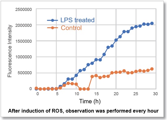
Time-lapse Imaging of ROS in LPS-treated Macrophages
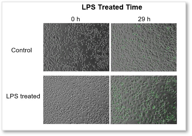
RAW264.7 macrophages were stained with ROS Assay Kit -Photo-oxidation Resistant DCFH-DA- and treated with lipopolysaccharide (LPS). Intracellular ROS fluorescence was monitored over time using fluorescence microscopy.
Results
- ROS fluorescence increased significantly after 10 hours of LPS treatment.
- Cell line
- RAW264.7
- Treatment
- LPS, 500 ng/ml.
- Imaging conditions
- Every hour for 30 hours.
GFP filter, Ex 450–490 nm, Em 500–550 nm.
Epifluorescence microscope, BZ-X800 (KEYENCE).
Products in Use
- Intracellular Total ROS
ROS Assay Kit -Photo-oxidation Resistant DCFH-DA-
References
- 1. Diokmetzidou and Scorrano (2025). Mitochondria–membranous organelle contacts at a glance . J Cell Sci DOI: 10.1242/jcs.263895
- 2. Rizzuto et al. (1998) Close Contacts with the Endoplasmic Reticulum as Determinants of Mitochondrial Ca2+ Responses. Science DOI: 10.1126/science.280.5370.1763
- 3. Rizzollo and Agostinis (2025). Mitochondria-Lysosome Contact Sites: Emerging Players in Cellular Homeostasis and Disease. Contact (Thousand Oaks) DOI: 10.1177/25152564251329250
- 4. Fransen et al. (2017). The Peroxisome-Mitochondria Connection: How and Why? Int J Mol Sci. DOI: 10.3390/ijms18061126
- 5. Digiovanni et al. (2025). ROS transfer at peroxisome-mitochondria contact regulates mitochondrial redox. Science DOI: 10.1126/science.adn2804
- 6. Kim and Lee (2024). Socialized mitochondria: mitonuclear crosstalk in stress. Exp Mol Med. DOI: 10.1038/s12276-024-01211-4.
- 7. Zorov et al. (2014). Mitochondrial Reactive Oxygen Species (ROS) and ROS-Induced ROS Release. Physiol Rev. DOI: 10.1152/physrev.00026.2013
- 8. Wang et al. (2025). Innate immune and metabolic signals induce mitochondria-dependent membrane lysis via mitoxyperiosis. Cell. DOI:10.1016/j.cell.2025.11.002
- 9. Cañeque et al. (2025). Activation of lysosomal iron triggers ferroptosis in cancer. Nature. DOI: 10.1038/s41586-025-08974-4
- 10. Saimoto et al. (2025). Lysosomal lipid peroxidation contributes to ferroptosis induction via lysosomal membrane permeabilization. Nat Commun. DOI: 10.1038/s41467-025-58909-w
- 11. Sassano et al. (2025). Endoplasmic reticulum–mitochondria contacts are prime hotspots of phospholipid peroxidation driving ferroptosis. Nature Cell Biology. DOI: 10.1038/s41556-025-01668-z