|
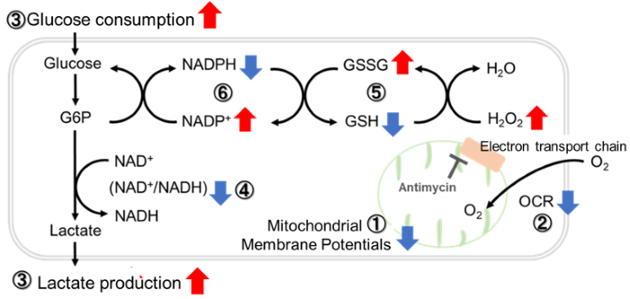
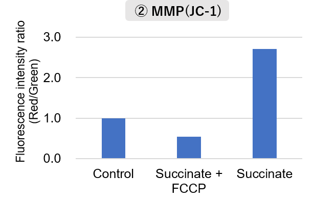
Mitochondrial function is generally evaluated using parameters such as oxygen consumption rate, membrane potential, ATP production, and reactive oxygen species (ROS). However, even when electron transport is maintained or enhanced, such changes do not necessarily contribute to normal cellular function. Recent studies have begun to reveal specific mechanisms by which alterations in mitochondrial electron transport support neurodegeneration or drug resistance in cancer cells.
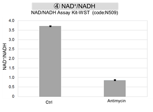
In the first study, tau, a microtubule-associated protein known for its role in axonal transport, was found to promote reverse electron transport (RET) through Complex I. Excessive RET increased ROS production and lowered the NAD⁺/NADH ratio, creating a vicious cycle that contributed to neurodegeneration. In the second study, colorectal cancer cells that survived chemotherapy exhibited increased mitochondrial biogenesis and oxidative metabolism, which supported drug resistance. Inhibition of Complex I further increased sensitivity to chemotherapy.
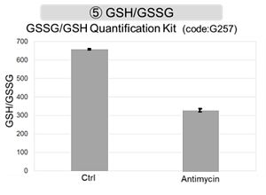
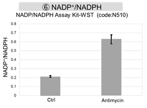
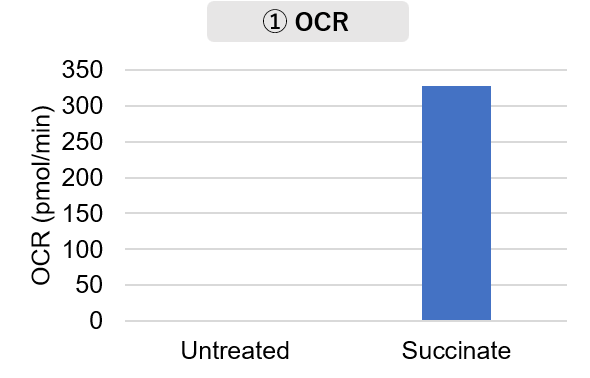
These findings suggest that maintained or enhanced mitochondrial electron transport and respiration do not necessarily indicate a beneficial cellular state. In addition to measuring Complex I activity and oxygen consumption rate (OCR), combining assessments of ATP production, mitochondrial ROS, and the NAD⁺/NADH ratio with cellular outcomes such as viability and drug sensitivity is important for understanding how mitochondrial alterations contribute to neuronal damage and chemotherapy resistance.
|
 |
Tau-induced mitochondrial reverse electron transport drives neurodegeneration
(Li et al., Neuron, 2026)
Summary:
Tau pathology and mitochondrial dysfunction are both associated with neurodegenerative diseases, but how tau influences mitochondrial electron transport has remained unclear. In this study, the authors examined tau's role in regulating RET across human iPSC-derived neurons, Drosophila, and mouse models of tauopathy, and found that tau directly interacts with a Complex I protein to regulate RET. Enhanced RET was associated with increased mitochondrial ROS, a reduced NAD⁺/NADH ratio, and neuronal injury-related phenotypes. These findings identify a previously unrecognized mechanism linking abnormal tau, RET, and neurodegeneration.
Highlighted technique:
To determine whether tau directly regulates reverse electron transport through Complex I, the authors compared RET- and FET-associated mitochondrial ROS production and NAD⁺/NADH status in isolated mitochondria. Tau localization, interaction with Complex I proteins, mitophagy-related markers, apoptosis-associated endpoints, and cell viability were examined to assess how RET-related metabolic changes are linked to neuronal injury.
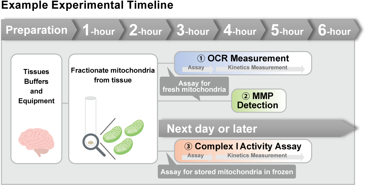
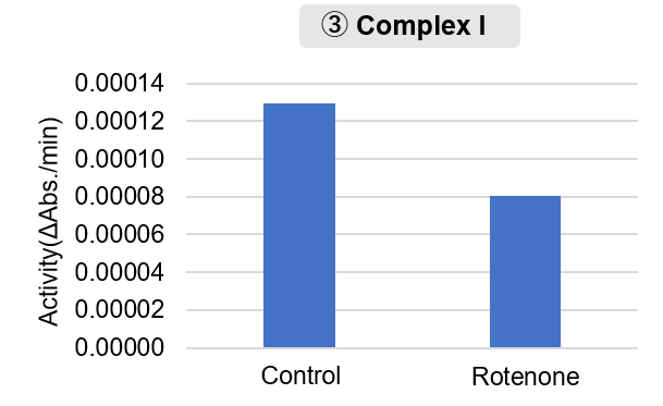
In addition to measuring Complex I activity and OCR after mitochondrial fractionation, mitochondrial ROS and intracellular NAD⁺/NADH measurements may help characterize metabolic shifts associated with RET.
Mitochondrial metabolism determines chemotherapy sensitivity in colorectal cancer
(Moss et al., Nature Metabolism, 2026)
Summary:
In this study, the authors evaluated mitochondrial abundance, Complex I activity, and oxidative metabolism in chemotherapy-surviving colorectal cancer cells, since the metabolic basis underlying their survival remained unclear. Surviving cells showed increased mitochondrial content, elevated Complex I activity, and enhanced oxidative metabolism, and Complex I inhibition restored chemotherapy sensitivity. These findings indicate that mitochondrial metabolic adaptation underlies chemotherapy resistance in colorectal cancer.
Highlighted technique:
To characterize metabolic features of chemotherapy-surviving colorectal cancer cells, the authors assessed mitochondrial abundance, Complex I activity in isolated mitochondria, and mitochondrial respiration. Complex I inhibition was then used to assess whether mitochondrial metabolic changes were associated with chemotherapy sensitivity.
OCR and ATP measurements in living cells detect changes in mitochondrial respiration and energy metabolism. Combining these with Complex I activity assays after mitochondrial fractionation identifies whether such changes are driven specifically by Complex I.
 
Mitochondrial Related Activity Indicators (click to open/close)
|