Mitochondria Research

Science Note

Lipid Homeostasis Controlled by Astrocytic OxPhos 

This article focusing on an intriguing study that emphasizes the critical role of astrocytic mitochondrial dysfunction in neurodegeneration and neuroinflammation, closely resembling Alzheimer's disease. The study underscores the significance of lipid homeostasis controlled by astrocytic OxPhos in maintaining brain health.     

Loss of fatty acid degradation by astrocytic mitochondria triggers neuroinflammation and neurodegeneration
Yashi Mi, et. al., Nature Metabolism (2023)

Point of Interest
- This study demonstrates that astrocytic OxPhos is crucial for breaking down fatty acids and maintaining lipid balance in the brain.
- When fatty acid levels overwhelm astrocytic OxPhos, this leads to lipid droplet buildup and neurodegeneration with similarities to Alzheimer's disease, including cognitive decline and demyelination.
- This process occurs as high acetyl-CoA levels trigger astrocyte reactivity through the activation of STAT3. 

Related Techniques
Total ROS detection ROS Assay Kit -Highly Sensitive DCFH-DA-
ROS Assay Kit -Photo-oxidation Resistant DCFH-DA-
Mitochondrial superoxide detection MitoBright ROS - Mitochondrial Superoxide Detection
Glycolysis/Oxidative phosphorylation Assay Glycolysis/OXPHOS Assay Kit
Glycolysis-related metabolites assay Glucose and Lactate ​Assay Kit
Oxygen consumption rate assay Extracellular OCR Plate Assay Kit
Mitochondrial membrane potential detection JC-1 MitoMP Detection Kit / MT-1 MitoMP Detection Kit
Intracellular lipid peroxide measurement Liperfluo
Lipid droplets detection and fatty acid uptake Lipi-Blue / Green / Red Deep Red, Fatty Acid Uptake Assay Kit
Related Applications

1. Monitoring ROS in Macrophage Phagocytosis

Dead cells (2&3) phagocytosed by Cell1 resulted in increased ROS(green).

ROS detection reagent allowed for reliable analysis of the role of ROS in phagocytosis. Its high intracellular residence and low background noise made it possible to perform long-term analysis of ROS production in the cell. This information can provide important insights into the mechanisms of phagocytosis and contribute to the development of treatments for diseases associated with macrophage dysfunction.

  > for detail experimental notes are available at Nikon web site.

Products in Use
   - ROS Assay Kit -Photo-oxidation Resistant DCFH-DA-

Related Product
  - ROS Assay Kit -Highly Sensitive DCFH-DA-


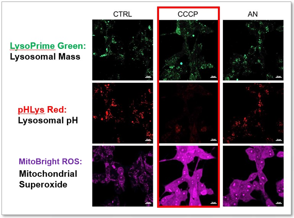
2. Mitochondrial Superoxide Detection in Senescent Cells

Background fluorescence caused by lipofuscin can be minimized by using a better fluorescent probe, as tested in TIG-1 cells. 

Lipofuscin accumulates in senescent cells, causing increased background fluorescence during observation. To minimize the effects of endogenous fluorescence from lipofuscin and other substances, a better fluorescent probe was tested in TIG-1 cells. Company T's product exhibited endogenous fluorescence, while MitoBright ROS Deep Red showed less background fluorescence. Researchers should compare sensitivity, wavelength, and channels and select the appropriate fluorescent probe to minimize endogenous fluorescence for accurate cellular senescence research.

Products in Use
  - MitoBright ROS - Mitochondrial Superoxide Detection


Mitochondria Research

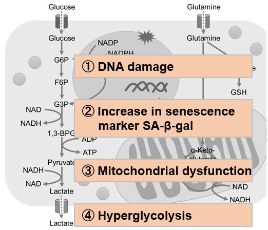
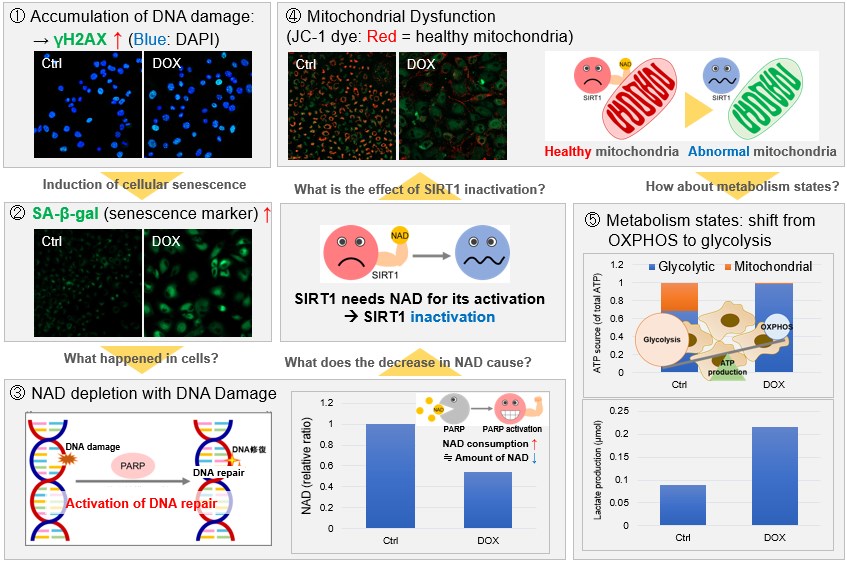
Mitochondria, which have important functions in cells, are not only a place for ATP production by oxidative phosphorylation, but their activity and dysfunction are closely related to cancer, senescence, and neurodegenerative diseases such as Alzheimer's disease and Parkinson's disease. Therefore, understanding the state of mitochondria is particularly important.


Mitophagy

Target Mitophagy
Reagent Mitophagy Detection Kit Keima-Red
Priciple Mtphagy Dye (included in Mitophagy Detection Kit) is a pH-sensitive fluorescent probe that accumulates in mitochondria and emits red fluorescence due to acidic conditions in a lysosome. A pH-sensitive ratiometric fluorescent protein. The excitation spectrum changes according to pH. This protein shows high fluorescence ratio (Ex. 550 nm/440 nm) values in a lysosome.
Accumulating in mitochondria Yes
(Covalent bond)
-
(Expression protein)
Live-cell staining
(Fixation after live-cell staining)

(-)

(-)
Fixed cell staining - -
Ex/Em (nm) 530/700  550 & 440/620
Staining time*1 30 min~ *2
Product code MD01 -

*1  Suitable staining time depends on sample type and reagent concentration.
*2  It is necessary to construct a keima expression strain.

Oxidative Stress

  Target Lipophilic Peroxide ROS Singlet oxygen Superoxide
Reagent MitoPeDPP ROS Assay Kit Si-DMA MitoBright ROS
Deep Red
MitoSOX Red
Principle A cell-permeant fluorescent probe that accumulates in mitochondria and specifically reacts with lipophilic peroxides in mitochondria to emit fluorescence. A cell-permeant fluorescent probe that  reacts with ROS  generated in mitochondria to emit fluorescence. A cell-permeant fluorescent probe that accumulates in mitochondria and specifically reacts with singlet oxigen generated in mitochondria to emit fluorescence. A cell-permeant fluorescent probe that accumulates in mitochondria and reacts with superoxide generated in mitochondria to emit fluorescence. A cell-permeant fluorescent probe that accumulates in mitochondria and reacts with superoxide generated in mitochondria to emit fluorescence.
Accumulating in mitochondria Yes
(Electrostatic interaction)
- Yes
(Electrostatic interaction)
Yes
(Electrostatic interaction)
Yes
(Electrostatic interaction)
Live-cell staining
(Fixed Cell staining)

(-)

(-)

(-)

(-)

(-)
Fixed cell staining - - - - -
Ex/Em(nm) 452/470 505/525 644/670 540/670 510/580
Common filter set FITC FITC Cy5 TexasRed
Cy5
TRITC
Staining time*1 30 min~ 15 min~ 45 min~ 10 min~ 10 min~
Product Code M466 R252 MT05 MT16 -

*1  Suitable staining time depends on sample type and reagent concentration.

Mitochondrial Membrane Potential

Target Mitochondrial Membrane Potential
Reagent MT-1 JC-1
Principle Cell-permeant fluorescent dyes accumulate in intact mitochondria due to the membrane potential. MT-1 is extremely photostable and more sensitive than JC-1 and can provide equivalent detection sensitivity to TMRE. A cell-permeant ratiometric fluorescent dye that accumulates in intact mitochondria due to the membrane potential. JC-1 forms aggregate (in healthy mitochondria) with red fluorescence.
As membrane potential decreases, JC-1 becomes monomers, which shows in green fluorescence.
Live-cell staining
(PFA fixation after staing)

(✓)

(-)
Fixed cell staining - -
Ex/Em(nm) 530-560/570-640 Monomer: 514/529
J-aggregation: 585/590
Common filter set TRITC
TexasRed
Monomer: FITC
J-aggregation: TRITC; TexasRed
Staining time*1 30  min 10~60  min
Product Code MT13 MT09

*1  Suitable staining time depends on sample type and reagent concentration.

Mitochondria Staining

Target Localization of mitochondria
Reagent MitoBright LT series MitoBright IM Red MitoTracker series Rhodamine 123
Principle

Cell-permeant fluorescent dyes accumulate in intact mitochondria due to the membrane potential.

Cell-permeant fluorescent dyes accumulate in intact mitochondria due to the membrane potential.

Cell-permeant fluorescent dyes accumulate in intact mitochondria due to the membrane potential.

Cell-permeant fluorescent dyes accumulate in intact mitochondria due to the membrane potential.

Live-cell staining
(PFA fixation after staining)

(✓)

(✓)

(✓)

(-)
Fixed cell staining - - - -
Combination with immunostaining - Conditionally -
Long-term retention × × ×
Ex/Em (nm) Green: 493/508
Red: 547/563
Deep Red:643/663
549/566 490/516 ~ 644/665 507/529
Common filter set Green: FITC

TRITC

-

FITC

Red: TRITC
DEEP Red: Cy5
Staining time*1 10 min~ 30 min~ 15~45 min 15 min~
Product Code MT10  MT11  MT12 MT15 - R233

*1  Suitable staining time depends on the sample type and reagent concentration.

Metal Ion Detection

Target Calcium ion (Ca2+) Iron ion(Fe2+)
Reagent Rhod 2-AM Mito-FerroGreen
Principle A cell-permeant fluorescent probe that accumulates in mitochondria and specifically reacts with calcium ion in mitochondria to emit red fluorescence. A cell-permeant fluorescent probe that accumulates in mitochondria and specifically reacts with ferrous ion in mitochondria to emit green fluorescence.
Accumulating in mitochondria
(Electrostatic interaction)

(Electrostatic interaction)
Live-cell staining
(PFA fixation after staing)

(-)

(-)
Fixed cell staining - -
Ex/Em(nm) 553/576 505/535
Common filter set TRITC FITC
Staining time*1 30~60  min 30 min~
Product Code R002 M489

*1  Suitable staining time depends on the sample type and reagent concentration.

Product Classification

Product Classification