

Science Note
Mitochondrial unfolded protein response in neural stem cell aging
| This article focusing on the mitochondrial protein folding stress is particularly significant in activated neural stem cells (NSCs) and neural progenitor cells (NPCs) within the neurogenic niche. This stress increases with age, leading to dysregulated cell cycles and mitochondrial activity in activated NSCs and NPCs in the dentate gyrus. | |
|
The mitochondrial unfolded protein response regulates hippocampal neural stem cell aging Point of Interest |
|
| Related Techniques | |
| Glycolysis/Oxidative phosphorylation assay | Glycolysis/OXPHOS Assay Kit |
| Oxygen consumption rate assay | Extracellular OCR Plate Assay Kit |
| Mitochondrial membrane potential detection | JC-1 MitoMP Detection Kit / MT-1 MitoMP Detection Kit |
| Mitochondrial superoxide detection | MitoBright ROS - Mitochondrial Superoxide Detection |
| Mitophagy Detection | Mitophagy Detection Kit and Mtphagy Dye |
| Cellular senescence detection (Live cell imaging or FCM) | Cellular Senescence Detection Kit |
| Cellular senescence detection (Plate reader) | Cellular Senescence Plate Assay Kit |
| Lysosomal function assay | Lysosomal pH and mass detection Kit |
| Related Applications | |
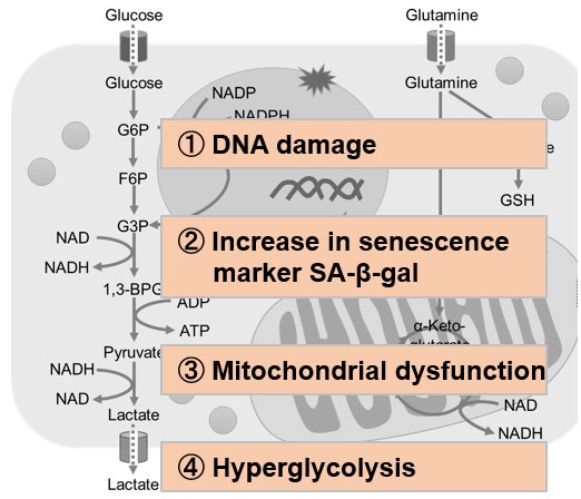
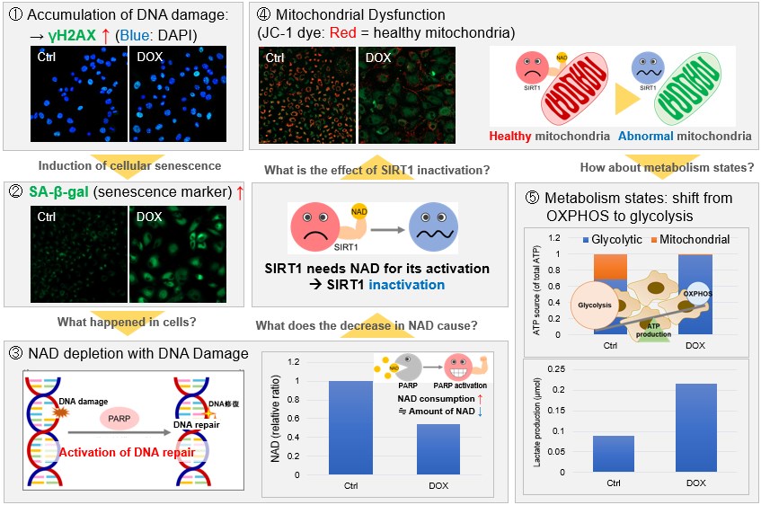
Metabolic shift to glycolysis in senescenct cells
Products in Use |
|
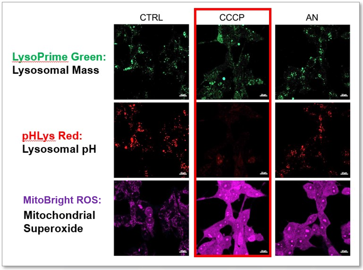
2. Simultaneously detection of Lysosomal and Mitochondrial Dysfunction
|
|
_20240723_en.jpg)
