|
Recent studies suggest that lipid metabolism is shaped not only by how fatty acids are broken down, but also by where lipids come from, how much enters the cell and where they are stored. In neurons, DDHD2, an enzyme that releases saturated fatty acids from stored intracellular lipids, supplied fuel for mitochondrial respiration and ATP production when glycolysis could not compensate. In acid-adapted glioblastoma cells, a sugar-rich surface layer limited extracellular lipid entry, while lipid droplets buffered incoming lipids and reduced lipid peroxidation and ferroptosis. Together, these studies show how lipid flux can support energy production or become a source of oxidative injury when cellular protective capacity is exceeded.
|
 |
DDHD2 provides a flux of saturated fatty acids for neuronal energy and function
(Saber et al., Nature Metabolism, 2025)
Summary:
In this study, cultured mouse neurons lacking DDHD2, an enzyme that releases fatty acids from stored cellular lipids, showed increased glycolysis but still had reduced mitochondrial respiration and intracellular ATP levels. Supplying long-chain saturated fatty acids in a form that cells could use for metabolism restored energy production. The findings identify internally released fatty acids as an important neuronal fuel, particularly during increased energy demand.
Highlighted technique:
To test whether loss of an intracellular fatty acid supply shifts neuronal energy production away from mitochondrial respiration and toward glycolysis, the authors compared control and Ddhd2-deficient mouse neurons. Intracellular ATP was measured by luminescence, while oxygen consumption and extracellular acidification were quantified by metabolic flux analysis.
As a complementary experiment, fatty acid uptake can be measured alongside intracellular ATP, glycolysis/OXPHOS balance and mitochondrial oxidative stress to assess extracellular lipid use.
Tumour acidosis remodels the glycocalyx to control lipid scavenging and ferroptosis
(Bång-Rudenstam et al., Nature Cell Biology, 2026)
Summary:
In this study, glioblastoma cells adapted to acidic conditions by forming a sugar-rich surface layer that restricted extracellular lipid entry. Extracellular lipid availability also supported lipid-droplet formation. Disrupting both the surface barrier and lipid-droplet formation caused extensive lipid peroxidation and ferroptosis, an iron-dependent form of cell death, revealing coordinated control of lipid entry, storage and survival.
Highlighted technique:
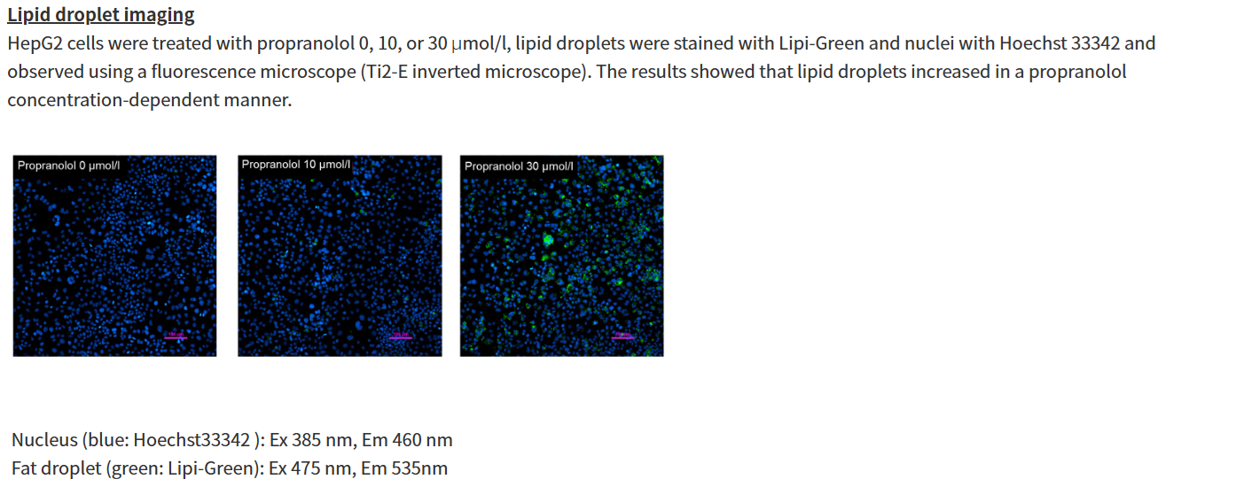
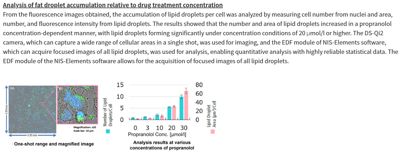
To evaluate whether restricted lipid entry and intracellular storage protect acid-adapted tumour cells from lipid overload, the authors measured fluorescent lipid-particle binding and uptake by flow cytometry and confocal imaging. Lipid droplets, lipid peroxidation and cytotoxicity were quantified by fluorescence-based assays, with selective inhibitors used to identify ferroptosis.
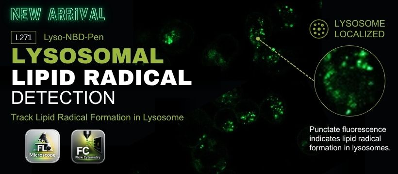
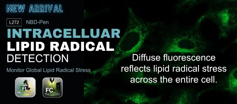
Track lipid droplets, early-stage intracellular lipid radicals, and early-stage lysosomal lipid radicals for a more complete understanding of lipotoxicity.
Lipid Metabolism Related Indicators (click to open/close)
|