Rethinking Glycolysis/OXPHOS in Cancer [June. 4, 2026]

Cancer cell metabolic states have recently been suggested to require multiparametric assessment, including glycolysis/OXPHOS dependence, mitochondrial abundance and membrane potential, and the utilization of metabolic substrates such as lactate. One study revealed that mitochondrial abundance does not necessarily correlate with OXPHOS dependence in CRC cells. Another showed that lactate is not merely a glycolytic waste product but a reusable mitochondrial fuel that can support OXPHOS and stemness in metastatic breast cancer cells.

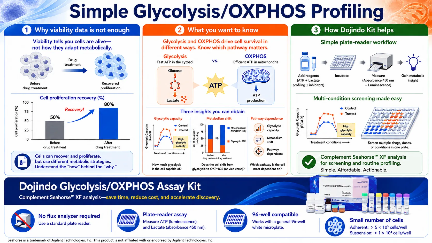
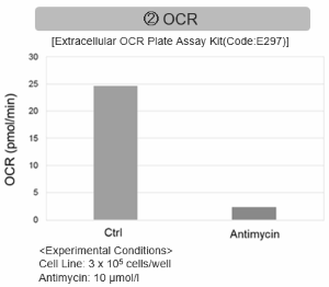
Mitochondrial, metabolic and bioenergetic adaptations drive plasticity of colorectal cancer cells and shape their chemosensitivity (Markov, et al., Cell Death & Disease, 2025)
Summary
This study demonstrates that cancer cell chemosensitivity cannot be understood solely through glycolytic or OXPHOS dependence. Using eight CRC cell lines, the authors showed that mitochondrial abundance does not necessarily reflect mitochondrial function or metabolic reliance. They further linked mitochondrial quantity, protein density, membrane potential, bioenergetic profiles, and metabolite patterns to drug-specific responses.  

Highlighted technique
To functionally classify CRC cell metabolism, the authors quantified mitochondrial membrane potential and mitochondrial ROS by flow cytometry, and measured oxygen consumption and extracellular acidification using Seahorse flux analysis to evaluate respiratory capacity, glycolytic activity, and their balance.

A Glycolysis/OXPHOS assay kit and OCR plate-assay kit provides a low-cost, low-cell-number approach screening for samples before committing to full Seahorse analysis.

Lactate mitochondrial oxidation drives stemness potential in metastatic breast cancer (Zhang, et al., Nature communications, 2025)
Summary
In this study, lactate is shown to be not merely a glycolytic byproduct but a mitochondrial fuel source in breast cancer cells undergoing metastasis. In detached cells that survive after loss of adhesion, lactate uptake and oxidation through the CD147/MCT1/LDHB complex support TCA metabolism, OXPHOS, and α-KG-DNMT3B-SOX2-associated stemness potential, linking lactate utilization to metastatic adaptation.

Highlighted technique
To examine whether lactate serves as a mitochondrial fuel linked to stemness potential, the authors combined isotope tracing of lactate-derived TCA metabolites with measurements of ATP production, Seahorse-based OCR, and Complex I activity. These metabolic readouts were integrated with tumorsphere formation and SOX2 expression to assess whether lactate oxidation supports OXPHOS and stemness-related sphere-forming capacity.

A mitochondrial fractionation kit enables the collection of intact mitochondria from tissue samples, allowing direct assessment of mitochondrial function, including OCR and Complex I activity. Additionally, intracellular ATP and α-KG can serve as readouts of mitochondrial metabolic changes.

Metabolic and Mitochondrial Activity Indicators (click to open/close)
Target Kit & Probes
Glycolysis/Oxidative phosphorylation Assay Glycolysis/OXPHOS Assay Kit
Oxygen consumption rate assay Extracellular OCR Plate Assay Kit
Intracellular ATP mesurement ATP Assay Kit-Luminescence
Extracellular ATP mesurement Extracellular ATP Assay Kit-Luminescence
ATP/ ADP ratio mesurement ADP/ATP Ratio Assay
Lactic Acid Measurement Lactate Assay Kit-WST
α-Ketoglutaric Acid Measurement α-Ketoglutarate Assay Kit-Fluorometric
Intact Mitochondria Fractionation IntactMito Fractionation Kit for Tissue
MitoComplex-I Activity Assay MitoComplex-I Activity Assay Kit
Mitochondrial Staining MitoBright LT Green / Red / Deep Red
Mitochondrial membrane potential detection JC-1 MitoMP Detection Kit, MT-1 MitoMP Detection Kit
Mitochondrial superoxide detection MitoBright ROS Deep Red - Mitochondrial Superoxide Detection
Application Note I (click to open/close)
  > Inhibition of Mitochondrial Electron Transport Chain

Antimycin stimulation of Jurkat cells was used to evaluate the changes in cellular state upon inhibition of the mitochondrial electron transport chain using a variety of indicators.

The results showed that inhibition of the electron transport chain resulted in (1) a decrease in mitochondrial membrane potential and (2) a decrease in OCR. In addition, (3) the NAD+/NADH ratio of the entire glycolytic pathway decreased due to increased metabolism of pyruvate to lactate to maintain the glycolytic pathway, (4) GSH depletion due to increased reactive oxygen species (ROS), and (6) increase in the NADP+/NADPH ratio due to decreased NADH required for glutathione biosynthesis were observed.


 Application Note II  (click to open/close)
  > Activity Evaluation of Mitochondria Fractionated from Mouse Brain


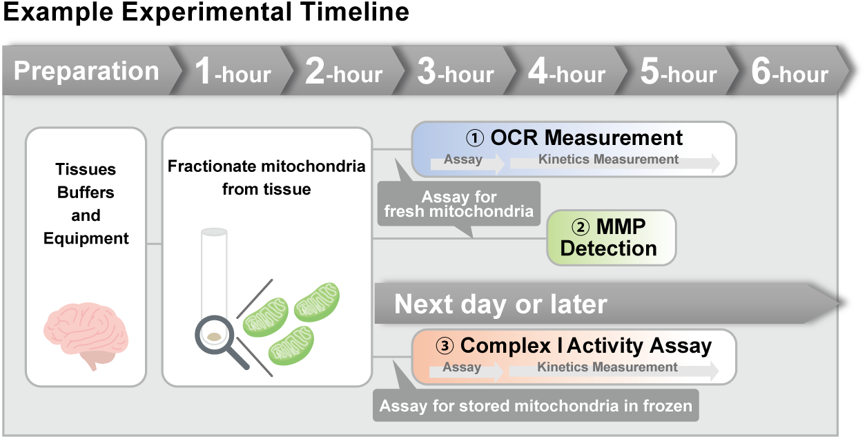
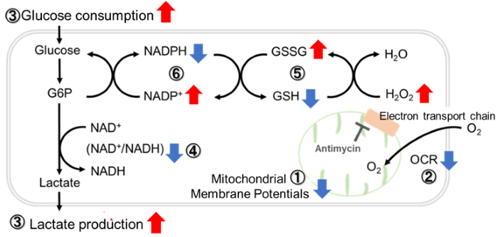
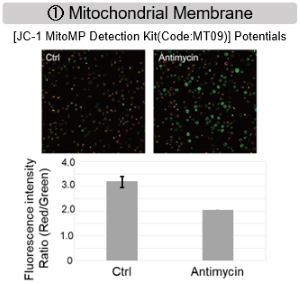
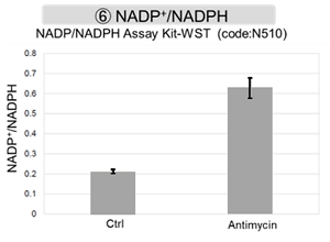
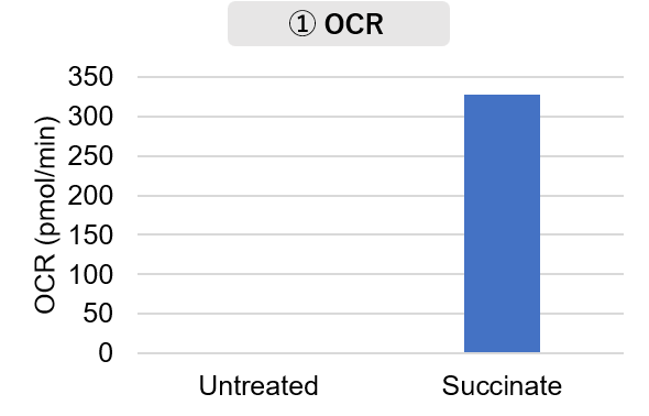
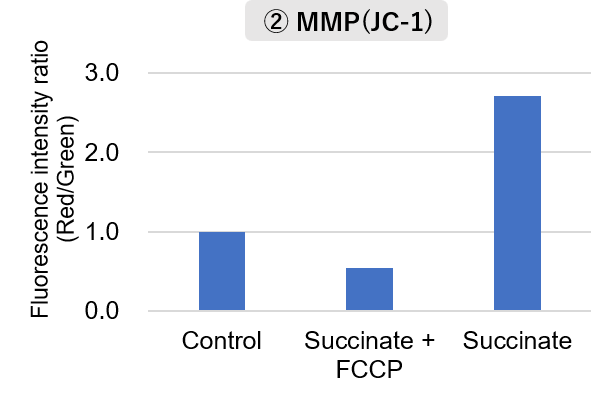
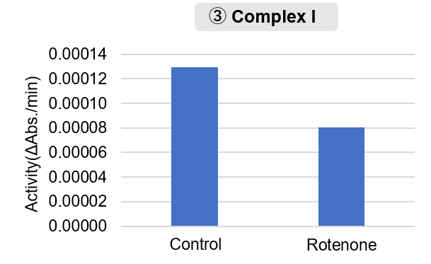
Mitochondria were isolated from mouse brain tissue, and oxygen consumption rate (OCR), mitochondrial membrane potential (MMP), and Complex I activity were measured.

The results showed that the addition of succinate, a substrate that activates Complex II of the electron transport chain, increased both OCR and MMP. In contrast, FCCP treatment reduced MMP, indicating that intact mitochondria were successfully fractionated.
Furthermore, in the Complex I activity assay, a decrease in activity was observed following treatment with rotenone, a Complex I inhibitor.

<Product used>
    Mitochondrial Fractionation: IntactMito Fractionation Kit for Tissue (Code: MT17)
 OCR measurement: Extracellular OCR Plate Assay Kit (Code: E297)
 MMP detection: JC-1 MitoMP Detection Kit (Code: MT09)
 Complex I activity assay:  MitoComplex- I Activity Assay Kit (Code: MT18)

<Experimental Conditions>
OCR Measurement   
    Amount of mitochondria: 50 μg/well (as protein levels)
    Succinate: 10 mmol/l
MMP Detection  
    Amount of mitochondria: 50 μg/well (as protein levels)
    Succinate: 10 mmol/l,   FCCP: 4 μmol/l
Complex I Activity Assay  
    Amount of mitochondria: 20 μg/well (as protein levels)
       Rotenone: 10 μmol/l

Product Classification

Product Classification