Metabolic Remodeling: How Cancer Survives [Oct. 15, 2025] 

Previous Science Note

Cancer has long been defined by the Warburg effect, yet many tumors also depend on mitochondrial OXPHOS, using both pathways as conditions change. This metabolic plasticity serves as a survival strategy that supports growth and stress tolerance under nutrient and oxygen constraints. To frame this landscape, we introduce a recent review that charts glycolysis, OXPHOS, glutaminolysis, and lipid metabolism in cancer and outlines actionable metabolic targets. We also highlight new research showing that cancers can reprogram the metabolism of neighbouring cells, notably shifting the glycolysis/OXPHOS balance in T cells via mitochondrial transfer and thereby weakening antitumor immunity.

Altered metabolism in cancer: insights into energy pathways and therapeutic targets (Molecular Cancer, 2024)
Summary: Tumor cells undergo metabolic reprogramming, switching between Glycolysis and OXPHOS in response to nutrient and oxygen constraints in the tumor microenvironment to sustain proliferation and stress tolerance.
This review integrates Glycolysis and OXPHOS with glutaminolysis and lipid metabolism in cancer, catalogues actionable targets from glucose transporters, glycolytic enzymes and LDH to electron transport and ATP synthesis, glutaminase, lipid synthesis and fatty-acid oxidation, and emphasizes metabolic plasticity and context-dependent drug sensitivity. Accordingly, analyzing Glycolysis/OXPHOS balance and switching with glucose uptake and lactate production, OCR, and mitochondrial function metrics is essential for revealing plasticity and context-specific vulnerabilities for combination and synthetic-lethal therapy.

 Related techniques  Glycolysis/OXPHOS Assay, Glucose Uptake Plate Assay

Immune evasion through mitochondrial transfer in the tumour microenvironment (Nature, 2025)
Summary: Cancer cells transfer mtDNA-mutant mitochondria to tumor-infiltrating T cells, reprogramming T-cell metabolism and shifting the glycolysis/OXPHOS balance. This mitochondrial transfer drives T-cell dysfunction and senescence, weakening antitumor immunity and pointing to interventions that block mitochondria transfer or modulate mitophagy.

Highlighted technique: In this study, co-cultured cancer cells and T cells, tracked fluorescently labeled mitochondria by confocal, time-lapse, and electron microscopy, and quantified transfer with flow cytometry. 

 Related techniques   Long-lasting Mitochondrial Staining, Mitophagy Detection

Previous Science Note

Related Techniques (click to open/close)
Target Kit & Probes
Glycolysis/oxidative phosphorylation assay Glycolysis/OXPHOS Assay Kit
Glucose Uptake Capacity Assay  Glucose Uptake Plate Assay Kit (NEW)
Glucose Uptake Assay Kit-Blue / Green / Red
Oxygen consumption rate assay Extracellular OCR Plate Assay Kit
Mitochondrial Staining MitoBright LT Green / Red / Deep Red
Mitochondrial membrane potential detection JC-1 MitoMP Detection Kit, MT-1 MitoMP Detection Kit
Mitophagy detection without trasfection Mitophagy Detection Kit
Cellular senescence detection SPiDER-βGal for live-cell imaging or flow cytometry / microplate reader / tissue samples.
SPiDER Blue: Fixed-cell multicolor senescence stain
ROS detection ROS Assay Kit -Highly Sensitive DCFH-DA- and ROS Assay Kit -Photo-oxidation Resistant DCFH-DA-
Apoptosis detection in multiple samples Annexin V Apoptosis Plate Assay Kit
Cell proliferation/ cytotoxicity assay Cell Counting Kit-8 and Cytotoxicity LDH Assay Kit-WST
Application Note (click to open/close)
  > Modulation of Glucose Uptake by Cytochalasin B and Insulin

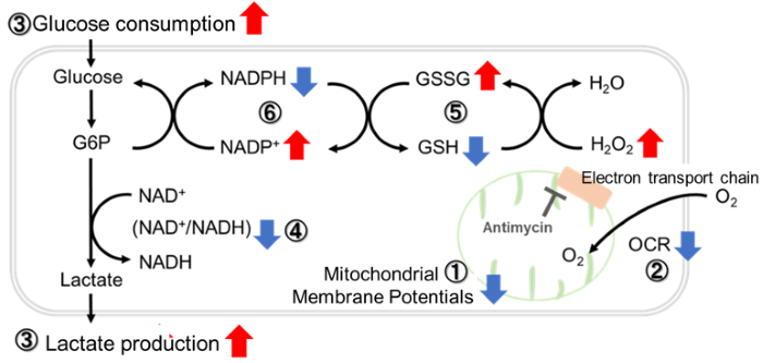
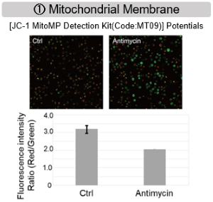
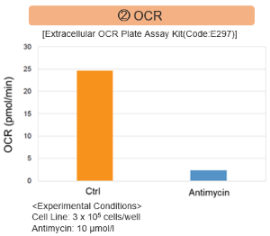
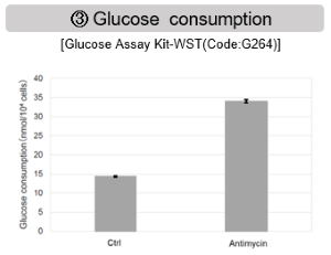
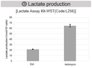
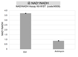
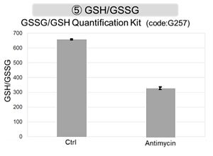
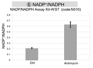
Antimycin stimulation of Jurkat cells was used to evaluate the changes in cellular state upon inhibition of the mitochondrial electron transport chain using a variety of indicators.

The results showed that inhibition of the electron transport chain resulted in (1) a decrease in mitochondrial membrane potential and (2) a decrease in OCR. In addition, (3) the NAD+/NADH ratio of the entire glycolytic pathway decreased due to increased metabolism of pyruvate to lactate to maintain the glycolytic pathway, (4) GSH depletion due to increased reactive oxygen species (ROS), and (6) increase in the NADP+/NADPH ratio due to decreased NADPH required for glutathione biosynthesis were observed. 

Products in Use
① JC-1 MitoMP Detection Kit
② Extracellular OCR Plate Assay Kit
③ Glucose Assay Kit-WST / Lactate Assay Kit-WST
④ NAD/NADH
⑤ GSSG/GSH Quantification Kit
⑥ NADP/NADPH Assay Kit


Previous Science Note  


Product Classification

Product Classification