Lysosome Damage Responses in Disease Biology [Jun. 16, 2026]


Lysosomes regulate degradation, membrane integrity, and immune-related processing. Lysosome damage responses are important for understanding how organelle injury influences disease-relevant cellular outcomes. Recent studies identified Parkinson's disease-associated VPS13C as an early factor recruited to damaged lysosomes, suggesting a membrane-protective response before severe rupture. In macrophages lacking TMEM175, lysosomal stress during tumor debris processing promoted inflammatory cytokine release and responses that help present tumor antigens to CD8⁺ T cells. These findings support lysosome damage responses as a relevant focus in disease research. 
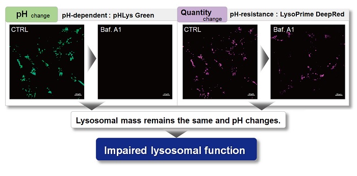
1. The bridge-like lipid transport protein VPS13C/PARK23 mediates ER-lysosome contacts following lysosome damage
(Nature Cell Biology, 2025)


Summary
This study showed that the Parkinson’s disease associated protein VPS13C acts as an early response factor to lysosome damage. VPS13C was recruited to damaged lysosomes within minutes and mediated ER lysosome contacts, suggesting a membrane protective response before severe rupture marked by Gal3. Its distinct timing from LRRK2, a Parkinson’s disease associated kinase recruited later to stressed lysosomes, suggests VPS13C dysfunction may reduce early lysosome damage resilience. 

Highlighted technique
To assess lysosome homeostasis associated with VPS13C loss, the authors compared wild type and VPS13C-knockout A549 cells. Lysosome-associated LAMP1 signal was evaluated by LAMP1 immunofluorescence, while lysosome acidification was measured using a pH-sensitive lysosome probe, providing readouts of lysosome status before damage-challenge experiments.

By combining pH-dependent and pH-independent lysosome probes, lysosomal pH and mass can be assessed by fluorescence imaging without immunostaining or transfection.

2. Deficiency of lysosomal TMEM175 in myeloid macrophages exerts anti-tumor immunity via inflammasome and cross-presentation pathway
(Nature Communications, 2026)


Summary
TMEM175 deficiency in macrophages showed that lysosomal dysfunction can convert tumor cell debris processing into an anti-tumor immune signal. After debris uptake, increased lysosomal membrane permeabilization and cathepsin B leakage activated NLRP3 inflammasome signaling and IL-1β and IL-18 secretion. Delayed lysosomal antigen degradation likely supported enhanced cross-presentation, strengthening CD8⁺ T cell immunity. These findings suggest that lysosomal stress in macrophages supports tumor antigen presentation and CD8⁺ T cell-mediated immune surveillance.

Highlighted technique:
To examine whether lysosomal dysfunction promotes inflammasome activation after tumor debris uptake, the authors exposed macrophages to B16-F10 tumor cell debris. They assessed lysosomal membrane damage by electron microscopy and cathepsin B immunofluorescence, measured Ca²⁺ changes and intracellular ROS with fluorescent probes, and evaluated NLRP3 activation by cytokine release and protein interaction assays.

Evaluating Ca²⁺ dynamics and intracellular ROS helps clarify lysosome-associated inflammasome activation. As a complementary approach, detecting intracellular and lysosomal lipid radicals may provide additional information on oxidative membrane stress associated with lysosomal dysfunction. 


All Related Techniques (click to open/close)
Target Kit & Probes
Lysosomal Function Analysis Kit Lysosomal Acidic pH Detection Kit - Green/Red and Green/Deep Red
High Specific Lysosommal Detection LysoPrime Green / Deep Red
Lysosomal Acidic pH Detection pHLys Red
Intracellular Calcium detection Calcium Kit II - Fluo 4 and Calcium Kit II - Fura 2
Total ROS detection Highly sensitive DCFH-DA or Photo-oxidation Resistant DCFH-DA
Intracellular lipid radical detection Lipid Radical Probe -NBD-Pen-
Lysosomal lipid radical detection Lysosomal Lipid Radical Probe -Lyso-NBD-Pen-
Lipid Peroxidation Assay Lipid Peroxidation Probe -BDP 581/591 C11-
Application Note (click to open/close)
  > Accurate Measurement for Lysosomal pH changes

With existing reagents, it was difficult to determine whether lysosomal mass or their function (pH) fluctuated because the discussion was based on changes in the fluorescence brightness of a single dye. This kit contains pHLys Green, which is highly specific to lysosomes and shows pH-dependent changes in fluorescence, and pH-resistant LysoPrime Deep Red. Using these two dyes, lysosomal pH and volume of the same sample can be measured for a detailed analysis of lysosomal function.

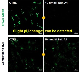
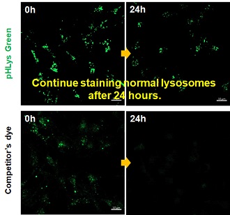
 Existing lysosomal pH detection reagents have issues with dye localization, pH sensitivity, and retention. pHLys Green is a dye that solves these issues. The improved dye retention and localization enable detection of normal lysosomes, and the improved pH sensitivity enables detection of slight pH changes.
1. High sensitive pH detection
Comparison of pH response of cells treated with low concentrations of lysosomal acidification inhibitor Bafilomycin A1
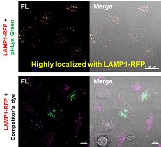
2. High specificity for lysosomes
Comparison of specificity for lysosomes using lysosomal marker protein LAMP1-GFP expressing cells
3. High retention in lysosomes
Comparison of intracellular retention

Product in Use:
   - Lysosomal Acidic pH Detection Kit-Green/Deep Red

Related Product:
   - Lysosomal Acidic pH Detection Kit-Green/Red
   - LysoPrime Deep Red - High Specificity and pH Resistance
   - pHLys Red - Lysosomal Acidic pH Detection


Product Classification

Product Classification