|
Lysosomes regulate intracellular degradation and membrane integrity, and recent studies have revealed their roles in ferroptosis by influencing lipid peroxidation and iron metabolism. These lysosomal functions are important for understanding how ferroptosis is controlled. One study showed that GPX4 inhibition causes heterogeneous ferroptotic death, with necrotic and apoptotic-like deaths occurring in parallel, and that lysosomal lipid peroxidation, rupture, and cathepsin involvement promote the switch toward propagating necrotic death. Another study identified NINJ2 as a LAMP1-interacting lysosomal factor that maintains membrane integrity, limits labile iron leakage, and suppresses ferroptosis sensitivity. These findings highlight lysosomal membrane integrity as a regulator of ferroptosis progression and susceptibility. |
||||||||||||||||||||||
|
Summary: This study shows that GPX4 inhibition-induced ferroptosis produces heterogeneous death profiles within cell populations, with necrotic and apoptotic-like deaths occurring in parallel, and that necrotic death is strongly associated with ferroptosis propagation. Mechanistically, ferroptotic stress induces lipid peroxidation at lysosomal membranes, leading to lysosome rupture and cathepsin involvement that promotes necrotic cell death and its spread to neighboring cells, highlighting the central role of lysosomes in ferroptosis propagation. Highlighted technique: To determine whether lysosome rupture is linked to necrotic ferroptosis, the authors used live-cell imaging with GFP-Galectin 3, a reporter that forms puncta when damaged lysosomes expose luminal glycans to the cytosol. They also used C11-BODIPY with lysosome staining dye to examine lipid peroxidation at lysosomal membranes, connecting lysosomal lipid peroxidation to lysosome rupture and necrotic ferroptosis propagation. |
||||||||||||||||||||||
|
Summary: This study reveals that NINJ2, originally identified as a nerve injury–induced cell adhesion molecule, also functions as a lysosome-associated regulator that interacts with LAMP1 and helps maintain lysosomal membrane integrity, thereby limiting the leakage of reactive labile iron and preserving ferritin stability. Loss of NINJ2 promotes lysosomal membrane permeabilization, ferritin degradation, and expansion of the labile iron pool, ultimately sensitizing cells to RSL3- and erastin-induced ferroptosis and linking lysosomal homeostasis to iron-dependent cell death. Highlighted technique: To examine whether loss of NINJ2 disrupts intracellular iron homeostasis, the authors treated control and NINJ2-knockout cells with ferric ammonium citrate as an iron source. They then measured intracellular iron levels using an iron assay kit and showed that NINJ2-deficient cells exhibited a marked increase in intracellular iron. |
||||||||||||||||||||||
|
Solutions for Lysosome Experiments |
||||||||||||||||||||||
All Related Techniques (click to open/close)
|
||||||||||||||||||||||
Application Note (click to open/close)
|
||||||||||||||||||||||
|
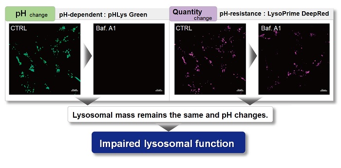
With existing reagents, it was difficult to determine whether lysosomal mass or their function (pH) fluctuated because the discussion was based on changes in the fluorescence brightness of a single dye. This kit contains pHLys Green, which is highly specific to lysosomes and shows pH-dependent changes in fluorescence, and pH-resistant LysoPrime Deep Red. Using these two dyes, lysosomal pH and volume of the same sample can be measured for a detailed analysis of lysosomal function.
|
|||
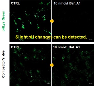
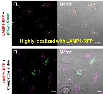
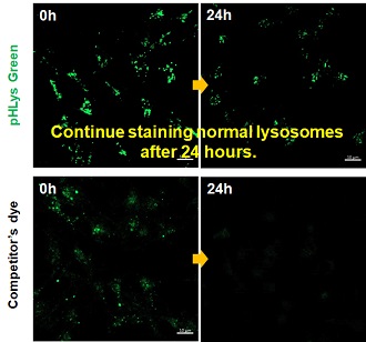
| Existing lysosomal pH detection reagents have issues with dye localization, pH sensitivity, and retention. pHLys Green is a dye that solves these issues. The improved dye retention and localization enable detection of normal lysosomes, and the improved pH sensitivity enables detection of slight pH changes. | |||
| 1. High sensitive pH detection Comparison of pH response of cells treated with low concentrations of lysosomal acidification inhibitor Bafilomycin A1 |
2. High specificity for lysosomes Comparison of specificity for lysosomes using lysosomal marker protein LAMP1-GFP expressing cells |
3. High retention in lysosomes Comparison of intracellular retention |
|
 |
 |
 |
|
|
Product in Use: Related Product: |
|||