


|
Summary: Combining lipid droplet staining with measurements of lipid peroxidation, intracellular lipid radicals, and lipid peroxides can provide a multidimensional assessment of the relationship between lipid droplet accumulation and cellular lipid oxidation.
Summary: Highlighted technique: Combining SA-β-gal detection with measurements of fatty acid uptake, lipid droplet accumulation, intracellular lipid radicals, lipid peroxides, and cell viability can provide a multidimensional view of how lipid storage and oxidative stress responses change during cellular senescence. Ferroptosis Related Indicators (click to open/close)
|

Application Note (click to open/close)
Experimental Example I: Hepatotoxicity test of drug-induced lipidosis using high-content imaging (click to open/close)
|
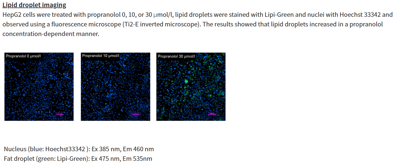
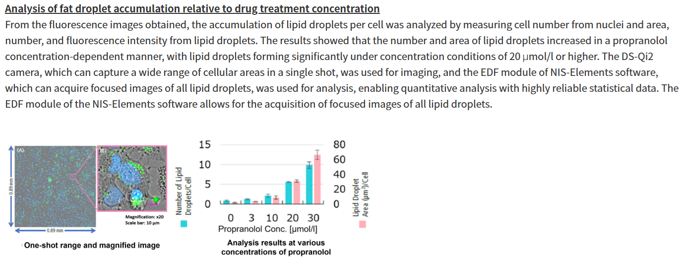
Propranolol (a sympathetic β-receptor blocker) was added to a human hepatocellular carcinoma cell line (HepG2 cells), and changes in lipid droplets were observed under a fluorescence microscope. The accumulation of lipid droplets was analyzed by measuring the number, area, and fluorescence intensity of lipid droplets from the acquired microscopic images.
For details of staining and analysis methods, please refer to "APPLICATION NOTE: Hepatotoxicity test of drug-induced lipidosis using high-content imaging" by Nikon Corporation. |
|
Experimental Example II: Changes in Each Indicator Following the Co-treatment of Ferroptosis Inducers and Lysosomal Inhibitors (click to open/close)
|
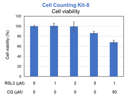
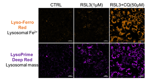
Previous studies have suggested that ferrotosis susceptibility varies among cancer cell lines. It has also been reported that increasing lysosomal stress in ferrotosis-resistant cancer cells can promote ferrotosis*. We treated A549 cells, which exhibit ferroptosis resistance, with the ferroptosis inducer RSL3 or RSL3 combined with the lysosomal inhibitor chloroquine (CQ) for 24 hours. Then, we analyzed changes in cell viability, lysosomal Fe²⁺, and lysosomal content. Treatment with RSL3 alone did not significantly alter cell viability or intracellular Fe²⁺ levels; however, some lysosomal aggregation (strong LysoPrime Deep Red signal) was observed. In contrast, cells treated with both RSL3 and CQ simultaneously exhibited increased intracellular Fe²⁺ levels, lysosomal enlargement, and decreased cell viability, which is consistent with previous reports. These results suggest that increased intracellular Fe²⁺ may promote ferroptosis. * Saimoto. Y, et al., Nature Communications, 2025, 16, 3554.
[Products in use] |
Experimental Example III: Comparison of Lysosomal Lipid Radical Detection in Ferroptosis-Sensitive and -Resistant Cells (click to open/close)
|
Changes in lysosomal lipid radicals were detected in HT-1080 cells, which are highly sensitive to ferroptosis, and A549 cells, which are resistant to ferroptosis, following treatment with RSL3, a ferroptosis inducer. In A549 cells, a ferroptosis-resistant cell line, no significant difference in fluorescence intensity was observed compared with the control, even when the RSL3 treatment concentration and duration of RSL3 exposure were increased (concentration: up to 2.5 μmol/L; duration: up to 3 hours). In contrast, in HT-1080 cells, a ferroptosis-sensitive cell line, treatment with 1 μmol/L RSL3 for 2 hours increased fluorescence derived from lysosomal lipid radicals and caused changes in their localization. These results demonstrate that this product can detect differences in lysosomal lipid radical generation and localization changes depending on the cell type. 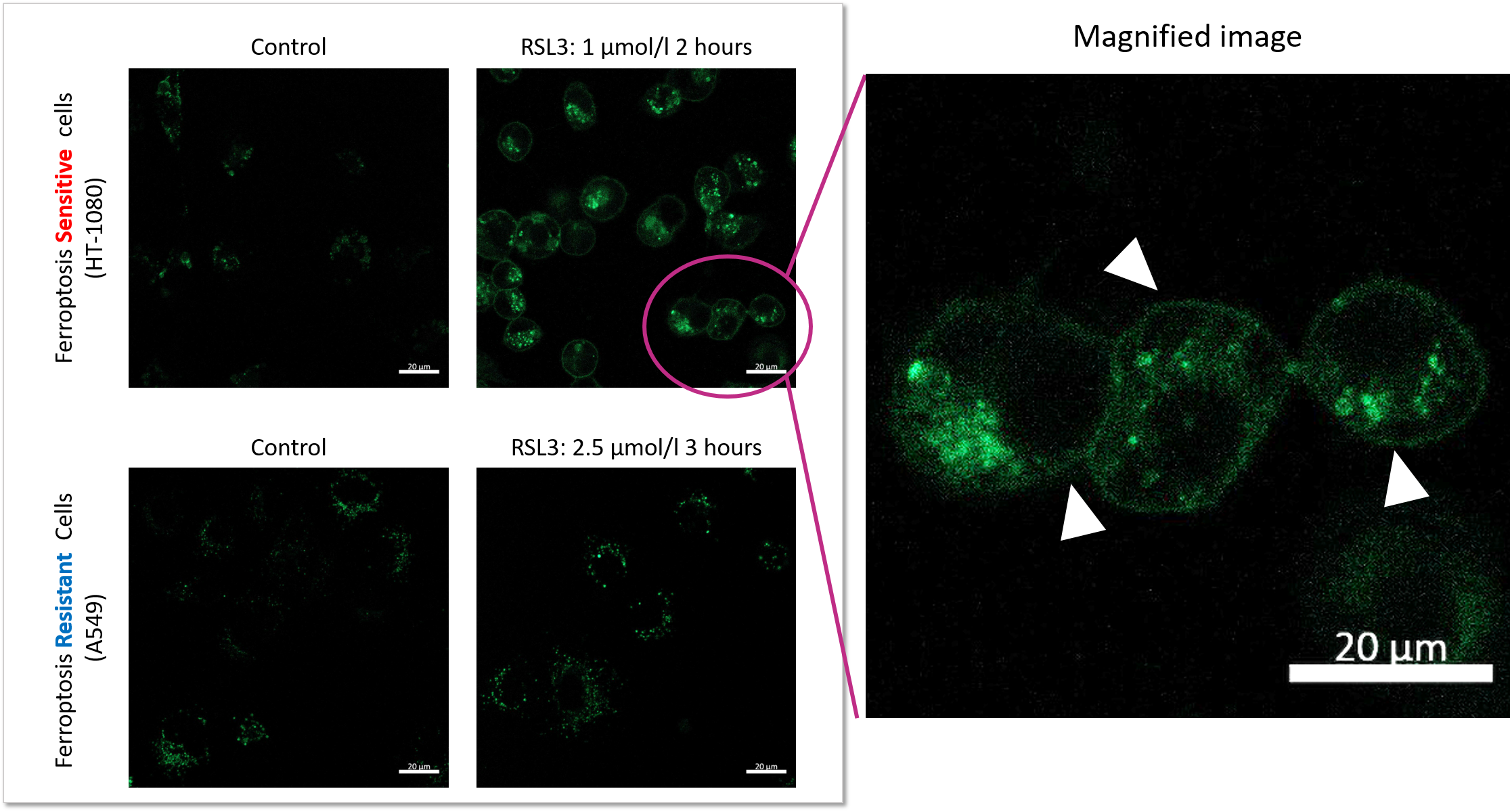 |