Emerging Lysosome Functions in Ferroptosis [Mar. 10, 2026]


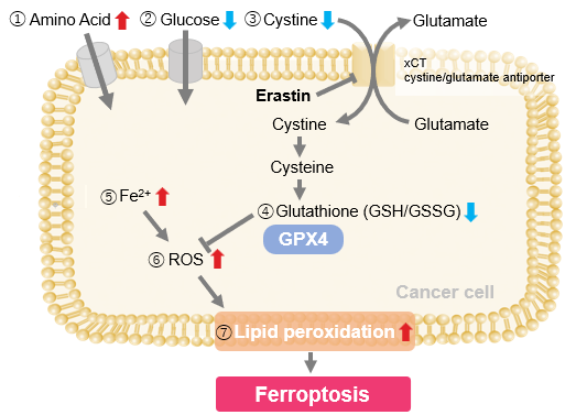
Ferroptosis is a form of cell death driven by lipid peroxidation, and its execution may be influenced by lysosomal function. Clarifying how lysosomes regulate ferroptosis sensitivity and cell death is important for understanding tumor responses and the control of cell death. Recent studies showed that, in non-small cell lung cancer, SMARCA4 deficiency confers ferroptosis resistance through a SMARCA4-ALDH16A1-TXN pathway involving lysosome mediated thioredoxin degradation. Another study showed that GPX4 inhibition gives rise to both apoptotic-like and necrotic-like cells during ferroptosis, and that lysosomal rupture, glutathione depletion, and cathepsin activity promote necrotic-like outcomes. These findings deepen our understanding of the relationship between lysosomes and ferroptosis.

1. Targeting ALDH16A1 mediated thioredoxin lysosomal degradation to enhance ferroptosis susceptibility in SMARCA4-deficient NSCLC (Nature Communications, 2025)
Summary: SMARCA4 deficiency, which has been recognized as a poor prognostic factor in non-small cell lung cancer, was shown to confer ferroptosis resistance. This study identified a novel SMARCA4–ALDH16A1–TXN regulatory pathway in which SMARCA4 induces ALDH16A1, thereby increasing ferroptosis susceptibility through lysosomal degradation and functional inhibition of the antioxidant factor thioredoxin.

Highlighted technique: To determine whether ALDH16A1 promotes lysosomal degradation of thioredoxin (TXN), the authors first examined whether TXN is regulated at the RNA or protein level in cells with altered ALDH16A1 expression. They then blocked new protein synthesis and added chloroquine or MG132 to test whether TXN is degraded through the lysosomal or proteasomal pathway. Finally, they visualized TXN and the lysosomal marker LAMP1 by immunofluorescence to assess whether ALDH16A1 loss or restoration changes TXN–lysosome colocalization.

2. Ferroptosis induces heterogeneous death profiles that are controlled by lysosome rupture (Developmental Cell, 2026)
Summary: Ferroptosis is a form of cell death driven by lipid peroxidation, and this study showed that GPX4 inhibition gives rise, within the same treated population, to cells with necrotic features and others with apoptotic features. Importantly, the study identified lysosomal rupture as a key factor that shifts this response toward necrotic outcomes, with glutathione depletion enhancing this shift and cathepsin activity contributing to necrotic cell rupture.

Highlighted technique: To determine whether lysosomal rupture occurs during GPX4 inhibition-induced cell death, the authors used cells expressing the fluorescent reporter GFP-Gal3 and monitored punctate signals that appear upon lysosomal damage. By tracking GFP-Gal3 together with Sytox staining after GPX4 inhibition, they examined whether lysosomal rupture occurs before plasma membrane rupture and whether it differs between cells showing necrotic or apoptotic features.

Ferroptosis Indicators (click to open/close)
Target Kit & Probes
Ferroptosis Indicator: ferrous ion (Fe2+) FerroOrange(intracellular), Mito-FerroGreen(mitochondria)
Ferroptosis Indicator: lipid peroxidation Liperfluo(intracellular), MitoPeDPP(mitochondria)
Lipid Peroxidation Assay Lipid Peroxidation Probe -BDP 581/591 C11-
Malondialdehyde Detection MDA Assay Kit
Lysosomal ferrous ion (Fe2+) detection Lyso-FerroRed
Lysosomal Function Analysis Kit Lysosomal Acidic pH Detection Kit -Green/Red and Green/Deep Red
High Specific Lysosommal Detection LysoPrime Green / Deep Red
Lysosomal Acidic pH Detection pHLys Red
Mitochondrial superoxide detection MitoBright ROS Deep Red - Mitochondrial Superoxide Detection
Total ROS detection Highly sensitive DCFH-DA or Photo-oxidation Resistant DCFH-DA
Glutathione Quantification GSSG/GSH Quantification Kit
Cystine Uptake detection Cystine Uptake Assay Kit
Application Note (click to open/close)
  > When Lysosomes Go Neutral: Iron Loss Unveiled

We investigated the transition of cellular metabolisms in A549 cells treated with erastin, a known ferroptosis inducer. Our results revealed the following.

Results
- The inhibition of cystine uptake by erastin led to a depletion of cysteine, which in turn increased the compensatory uptake of other amino acids.
- Glucose uptake, which typically promotes ferroptosis*, was found to decrease upon erastin treatment, suggesting a potential cellular self-defense mechanism.
- The depletion of cysteine resulted in a decrease in glutathione levels and an increase in Fe2+, ROS, and lipid peroxides, all of which are recognized markers of ferroptosis.

  Cell Line: A549
  Incubation Conditions: 100 μmol/l Erastin/MEM, 37℃, 3h
  *Reference: Xinxin Song, et al., Cell Reports, (2021)

Products in Use
① Amino Acid Uptake: Amino Acid Uptake Assay Kit
② Glucose Uptake: Glucose Uptake Assay Kit-Green
③ Cystine Uptake: Cystine Uptake Assay Kit
④ Intracellular glutathione: GSSG/GSH Quantification Kit
⑤ Intracellular labile Fe: FerroOrange
⑥ Intracellular total ROS: ROS Assay Kit -Highly Sensitive DCFH-DA-
⑦ Lipid Peroxides: Liperfluo


Product Classification

Product Classification