New Insights into GPX4 Control of Ferroptosis [Jan. 6, 2026]


GPX4 is a central suppressor of ferroptosis that reduces peroxidized phospholipids within cellular membranes, and its regulation is closely linked to the prevention of neurodegenerative diseases and the development of cancer therapies. Accordingly, new insights into GPX4 biology are important for advancing our understanding of ferroptosis control. Recent studies have shown that mutations disrupting the membrane-anchoring mechanism of GPX4 cause its mislocalization, which is sufficient to induce ferroptosis and neurodegeneration independently of enzymatic activity. In addition, PRDX6 has been identified as a selenium-acceptor protein required for GPX4 synthesis, and loss of PRDX6 leads to reduced GPX4 abundance and increased sensitivity to ferroptosis. Together, these findings provide an important foundation for modulating ferroptosis through GPX4 regulation and translating this knowledge into therapeutic applications.

1. A fin-loop-like structure in GPX4 underlies neuroprotection from ferroptosis (Cell, 2025)
Summary: GPX4 is a key enzyme in suppressing ferroptosis, but the importance of its subcellular localization has not been fully elucidated. Using GPX4 variants carrying mutations in the membrane-anchoring fin-loop, this study demonstrates across human iPSC-derived models, organoids, and mice that disruption of this anchoring mechanism is sufficient to trigger ferroptosis, and that GPX4 mislocalization rather than loss of enzymatic activity alone directly underlies ferroptosis-driven neurodegeneration.

Highlighted technique: To evaluate the importance of GPX4 localization, patient-derived primary fibroblasts carrying the R152H mutation in the membrane-anchoring fin-loop of GPX4 were used to analyze the impact of anchor disruption. Lipid peroxidation was assessed using BODIPY 581/591 C11, demonstrating that the R152H mutant exhibits increased accumulation of peroxidized lipids.

2. PRDX6 dictates ferroptosis sensitivity by directing cellular selenium utilization (Molecular Cell, 2024)
Summary: GPX4 is a selenium-dependent phospholipid hydroperoxide reductase and the central suppressor of ferroptosis, whose proper protein expression requires efficient selenium incorporation. Although PRDX6 itself does not directly suppress ferroptosis, this study demonstrates that PRDX6 acts as a selenium-acceptor protein essential for GPX4 synthesis, such that loss of PRDX6 reduces GPX4 abundance and increases ferroptosis sensitivity.

Highlighted technique: Using PRDX6 KO cells, this study shows that loss of PRDX6 markedly reduces GPX4 protein levels without altering GPX4 mRNA expression, demonstrating that PRDX6 is required for GPX4 synthesis rather than transcription. Consistently,PRDX6-deficient mice exhibit a selective reduction of GPX4 expression in the brain, where selenium supply and utilization are physiologically constrained, highlighting the essential role of PRDX6 in maintaining GPX4 abundance under selenium-limited conditions.

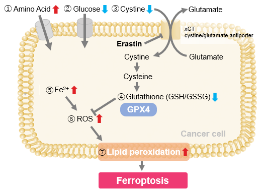
Ferroptosis Indicators (click to open/close)
Target Kit & Probes
Lysosomal ferrous ion (Fe2+) detection Lyso-FerroRed NEW
Ferroptosis Indicator: ferrous ion (Fe2+) FerroOrange(intracellular), Mito-FerroGreen(mitochondria)
Ferroptosis Indicator: lipid peroxidation Liperfluo(intracellular), MitoPeDPP(mitochondria)
Lipid Peroxidation Assay Lipid Peroxidation Probe -BDP 581/591 C11-
Malondialdehyde Detection MDA Assay Kit
Mitochondrial superoxide detection MitoBright ROS Deep Red - Mitochondrial Superoxide Detection
Total ROS detection Highly sensitive DCFH-DA or Photo-oxidation Resistant DCFH-DA
Glutathione Quantification GSSG/GSH Quantification Kit
Cystine Uptake detection Cystine Uptake Assay Kit
Application Note (click to open/close)
  > When Lysosomes Go Neutral: Iron Loss Unveiled

We investigated the transition of cellular metabolisms in A549 cells treated with erastin, a known ferroptosis inducer. Our results revealed the following.

Results
- The inhibition of cystine uptake by erastin led to a depletion of cysteine, which in turn increased the compensatory uptake of other amino acids.
- Glucose uptake, which typically promotes ferroptosis*, was found to decrease upon erastin treatment, suggesting a potential cellular self-defense mechanism.
- The depletion of cysteine resulted in a decrease in glutathione levels and an increase in Fe2+, ROS, and lipid peroxides, all of which are recognized markers of ferroptosis.

  Cell Line: A549
  Incubation Conditions: 100 μmol/l Erastin/MEM, 37℃, 3h
  *Reference: Xinxin Song, et al., Cell Reports, (2021)

Products in Use
① Amino Acid Uptake: Amino Acid Uptake Assay Kit
② Glucose Uptake: Glucose Uptake Assay Kit-Green
③ Cystine Uptake: Cystine Uptake Assay Kit
④ Intracellular glutathione: GSSG/GSH Quantification Kit
⑤ Intracellular labile Fe: FerroOrange
⑥ Intracellular total ROS: ROS Assay Kit -Highly Sensitive DCFH-DA-
⑦ Lipid Peroxides: Liperfluo


Product Classification

Product Classification