Lysosome Dynamics Shape Ferroptosis Sensitivity [Nov. 17, 2025]


Ferroptosis is an iron-dependent form of cell death shaped by lipid peroxidation as well as intracellular factors such as iron localization and lysosomal acidity. Lysosomes function as central sites for iron storage and mobilization, and emerging evidence suggests that iron activation and lipid peroxidation within lysosomes contribute to ferroptosis induction. Recent studies show that BDH2-dependent iron transfer between mitochondria and lysosomes in melanoma prevents Fe(II) sequestration, whose loss increases ferroptosis vulnerability. Another study demonstrates that lysosomal alkalinization during senescence causes abnormal Fe(II) retention, forming a major basis of ferroptosis resistance. These findings highlight lysosome-centered iron regulation as a key determinant of ferroptosis sensitivity.

1. BDH2-driven lysosome-to-mitochondria iron transfer shapes ferroptosis vulnerability of the melanoma cell states (Nature Metabolism, 2025)
Summary: Melanoma cells exist in a melanocytic (MEL) state and a drug-tolerant mesenchymal-like (MES) state. BDH2 mediates Fe2+  transfer at mitochondria–lysosome contacts in MEL cells and maintains lysosomal and mitochondrial homeostasis, and its loss in MES cells causes lysosomal iron sequestration and ferroptosis sensitivity, which can be reversed by restoring BDH2 or adding 2,5-DHBA, also increasing metastatic ability.

Highlighted technique: In this study, the authors aimed to determine how Fe2+ is distributed among intracellular organelles by performing live-cell imaging–based visualization and quantification of iron. They combined fluorescent probes to visualize Fe(II) within lysosomal regions and compare iron levels between MEL and MES cells, and further quantified mitochondrial iron using a mitochondria-specific probe to analyze iron compartmentalization across organelles.

 Related technique   Intracellular Fe2+ Detection, Mitochondrial Fe2+ Detection

2. Senescence-associated lysosomal dysfunction impairs cystine deprivation-induced lipid peroxidation and ferroptosis (Nature Communications, 2025)
Summary: This study showed that senescence impairs lysosomal acidification, leading to the abnormal retention of Fe2+ within lysosomes and thereby inhibiting cystine-deprivation-induced lipid peroxidation and ferroptosis. The authors also demonstrated that restoring lysosomal acidity through V-ATPase activation increases ferroptosis sensitivity in senescent cells.

Highlighted technique: This study aimed to clarify how lysosomal dysfunction in senescent cells alters intracellular Fe2+ distribution and ferroptosis sensitivity. To address this question, the authors used live-cell imaging with fluorescent probes to detect lysosomal Fe2+ and measure lysosomal pH, allowing them to evaluate iron sequestration and changes in intracellular iron distribution.

 Related technique   Lysosomal Fe2+ Detection, Lysosome function Analysis

Ferroptosis Indicators (click to open/close)
Target Kit & Probes
Lysosomal ferrous ion (Fe2+) detection Lyso-FerroRed NEW
Ferroptosis Indicator: ferrous ion (Fe2+) FerroOrange(intracellular), Mito-FerroGreen(mitochondria)
Ferroptosis Indicator: lipid peroxidation Liperfluo(intracellular), MitoPeDPP(mitochondria)
Lipid Peroxidation Assay Lipid Peroxidation Probe -BDP 581/591 C11-
Malondialdehyde Detection MDA Assay Kit
Mitochondrial superoxide detection MitoBright ROS Deep Red - Mitochondrial Superoxide Detection
Total ROS detection Highly sensitive DCFH-DA or Photo-oxidation Resistant DCFH-DA
Glutathione Quantification GSSG/GSH Quantification Kit
Cystine Uptake detection Cystine Uptake Assay Kit
Application Note (click to open/close)
  > When Lysosomes Go Neutral: Iron Loss Unveiled

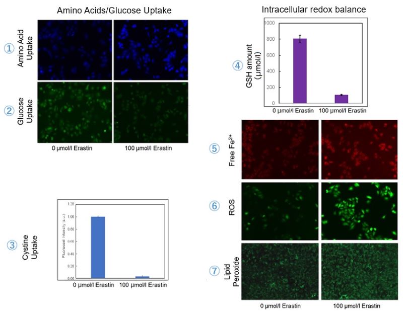
We investigated the transition of cellular metabolisms in A549 cells treated with erastin, a known ferroptosis inducer. Our results revealed the following.

Results
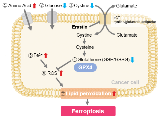
- The inhibition of cystine uptake by erastin led to a depletion of cysteine, which in turn increased the compensatory uptake of other amino acids.
- Glucose uptake, which typically promotes ferroptosis*, was found to decrease upon erastin treatment, suggesting a potential cellular self-defense mechanism.
- The depletion of cysteine resulted in a decrease in glutathione levels and an increase in Fe2+, ROS, and lipid peroxides, all of which are recognized markers of ferroptosis.

  Cell Line: A549
  Incubation Conditions: 100 μmol/l Erastin/MEM, 37℃, 3h
  *Reference: Xinxin Song, et al., Cell Reports, (2021)

Products in Use
① Amino Acid Uptake: Amino Acid Uptake Assay Kit
② Glucose Uptake: Glucose Uptake Assay Kit-Green
③ Cystine Uptake: Cystine Uptake Assay Kit
④ Intracellular glutathione: GSSG/GSH Quantification Kit
⑤ Intracellular labile Fe: FerroOrange
⑥ Intracellular total ROS: ROS Assay Kit -Highly Sensitive DCFH-DA-
⑦ Lipid Peroxides: Liperfluo


Product Classification

Product Classification