Lysosomal Control of Ferroptosis: Iron and Acidity as Key Determinants [Oct 7, 2025]


Recently, the importance of lysosomal iron in ferroptosis induction has attracted increasing attention. Recent studies in melanoma have identified a protein involved in iron flux at mitochondria–lysosome contact sites, revealing that organelle-to-organelle iron compartmentalization plays a role in ferroptosis regulation. In addition, research on senescent cells has highlighted that, despite iron accumulation, ferroptosis resistance is linked to lysosomal pH, demonstrating that lysosomal acidity is another key determinant of ferroptosis sensitivity. Together, these studies deepen our understanding of how the lysosomal environment contributes to ferroptosis regulation.

1. BDH2-driven lysosome-to-mitochondria iron transfer shapes ferroptosis vulnerability of the melanoma cell states (Nature Metabolism, 2025)
Summary: This study identifies how melanoma cells control iron inside the cell by focusing on the enzyme BDH2 at mitochondria–lysosome contact sites. It shows that BDH2 moves iron into mitochondria to keep lysosomes acidic, and when BDH2 is lost, iron builds up in lysosomes and the cells become more sensitive to ferroptosis.

Highlighted technique: To compare organelle iron distribution between melanoma cell states, the authors stained cells with FerroOrange for ferrous iron and lysosome detection probes. They observed increased overlap of both dyes in mesenchymal-like cells, indicating iron enrichment at mitochondria–lysosome contact regions during the phenotype transition.

 Related technique  Intracellular Fe2+ detection (as used in this article), pH-Insensitive Lysosome Detection

2. Senescence-associated lysosomal dysfunction impairs cystine deprivation-induced lipid peroxidation and ferroptosis (Nature Communications, 2025)
Summary: Recent studies emphasize the role of lysosomal iron in driving ferroptosis, but this study aimed to clarify why senescent cells remain resistant despite iron accumulation. The authors found that loss of lysosomal acidity in senescent cells traps ferrous iron and suppresses lipid peroxidation, whereas restoring acidity with the V-ATPase activator EN6 reactivated ferroptosis, revealing that both iron and lysosomal acidity are essential for ferroptosis regulation.

Highlighted technique: To test whether restoring lysosomal acidity can reverse ferroptosis resistance in senescent cells, EN6 was used to activate V-ATPase and reacidify lysosomes. Lysosomal pH, lysosomal ferrous ions, and lipid peroxidation were visualized, revealing that acidity restoration released trapped iron within lysosomes and reactivated ferroptosis.

 Related technique  Lysosome Acidic pH Detection, Cellular Senescence Detection

Related Techniques (click to open/close)
Target Kit & Probes
Ferroptosis Indicator: ferrous ion (Fe2+) FerroOrange(intracellular), Mito-FerroGreen(mitochondria)
Ferroptosis Indicator: lipid peroxidation Liperfluo(intracellular), MitoPeDPP(mitochondria)
Lipid Peroxidation Assay Lipid Peroxidation Probe -BDP 581/591 C11-
Lysosomal function Lysosomal Acidic pH Detection Kit -Green/Red and Green/Deep Red
Cellular senescence detection SPiDER-βGal for live-cell imaging or flow cytometry / microplate reader / tissue samples
Blue cellular senescence detection dye for fixed cells,  SPiDER Blue
Glycolysis/Oxidative phosphorylation Assay Glycolysis/OXPHOS Assay Kit
Oxygen consumption rate assay Extracellular OCR Plate Assay Kit
Total ROS detection Highly sensitive DCFH-DA or Photo-oxidation Resistant DCFH-DA
Apoptosis detection in multiple samples Annexin V Apoptosis Plate Assay Kit
Cell proliferation/ cytotoxicity assay Cell Counting Kit-8 and Cytotoxicity LDH Assay Kit-WST
Application Note (click to open/close)
  > When Lysosomes Go Neutral: Iron Loss Unveiled

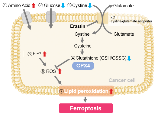
We investigated the transition of cellular metabolisms in A549 cells treated with erastin, a known ferroptosis inducer. Our results revealed the following.

Results
- The inhibition of cystine uptake by erastin led to a depletion of cysteine, which in turn increased the compensatory uptake of other amino acids.
- Glucose uptake, which typically promotes ferroptosis*, was found to decrease upon erastin treatment, suggesting a potential cellular self-defense mechanism.
- The depletion of cysteine resulted in a decrease in glutathione levels and an increase in Fe2+, ROS, and lipid peroxides, all of which are recognized markers of ferroptosis.

  Cell Line: A549
  Incubation Conditions: 100 μmol/l Erastin/MEM, 37℃, 3h
  *Reference: Xinxin Song, et al., Cell Reports, (2021)


Products in Use
① Amino Acid Uptake: Amino Acid Uptake Assay Kit
② Glucose Uptake: Glucose Uptake Assay Kit-Green
③ Cystine Uptake: Cystine Uptake Assay Kit
④ Intracellular glutathione: GSSG/GSH Quantification Kit
⑤ Intracellular labile Fe: FerroOrange
⑥ Intracellular total ROS: ROS Assay Kit -Highly Sensitive DCFH-DA-
⑦ Lipid Peroxides: Liperfluo


Product Classification

Product Classification